05 January 2021: Clinical Research
Utility of Magnetic Resonance Imaging in Diagnosis of Prenatal Non-Visualization of the Fetal Gallbladder: A Case-Series Study
Yuanhe Wang1ABCDEF, Jinling Zhou1C, Meixiang Deng2D, Xiaomiao Xiang3DF, Xiaojun Zhu1E*DOI: 10.12659/MSM.927474
Med Sci Monit 2021; 27:e927474


Abstract
BACKGROUND: This study aimed to assess the utility of magnetic resonance imaging (MRI) in the diagnosis of prenatal non-visualization of the fetal gallbladder (PNVGB).
MATERIAL AND METHODS: The clinical data of 32 pregnant women with PNVGB who underwent MRI examination during the second and third trimester of pregnancy were collected and their outcomes were analyzed.
RESULTS: MRI showed that 26 patients (81.3%) had isolated PNVGB and 6 (18.8%) had additional malformations. In 26 patients with isolated PNVGB, 7 were found in the gallbladder on MRI and 4 were found on subsequent ultrasonography. One patient had termination of pregnancy (TOP) and 1 patient was lost to follow-up; the remaining 24 patients were known to deliver a healthy child. Among the 6 patients with additional malformations, 3 terminated their pregnancies due to combined severe abnormalities: 1 patient with horseshoe kidney and 1 with fetal echogenic bowel both had a healthy child, while 1 with fetal growth restriction (FGR) delivered a child who walked on tiptoe.
CONCLUSIONS: MRI contributes to identifying PNVGB detected or suspected by ultrasonography.
Keywords: gallbladder diseases, Magnetic Resonance Imaging, Prenatal Diagnosis, Fetus, Gallbladder, Gestational Age, Pregnancy, Pregnancy Outcome
Background
Prenatal non-visualization of the fetal gallbladder (PNVGB) is uncommon in clinical practice, accounting for only about 0.10–0.15% of pregnancies [1,2]. Bile is formed by the fetal hepatic cells at 12 gestational weeks and enters the duodenum through the bile duct after 13 gestational weeks. The length, anteroposterior diameter, and transverse diameter of the fetal gallbladder have a linear relationship with gestational age between 15 weeks and 30 weeks, after which a plateau is observed [3,4]. The fetal gallbladder can be observed by transvaginal ultrasound at 14–16 weeks of gestation in 99.9% of pregnancies [1,2]. In 95% of pregnancies, it can be observed at 24–32 weeks of gestation by transabdominal ultrasound [5]. However, after 35 weeks, the gallbladder may not be visualized at the complete contraction due to the enhanced contraction function. Additionally, other factors like fetal positions and operating methods may also lead to PNVGB.
PNVGB is reported to have a correlation with benign conditions, including isolated gallbladder agenesis or several severe disorders such as cystic fibrosis, aneuploidies, and biliary atresia [6]. Gallbladder agenesis is a benign condition, with an incidence rate of 1/6300 [7], while biliary atresia is a severe disease associated with liver transplantation and death [8]. Typically, the gallbladder is very small or not seen by ultrasonography in cases of biliary atresia, and biliary atresia, which is characterized by intra- and extrahepatic bile duct obliteration, can progress rapidly to liver fibrosis and cirrhosis, and can even result in death if untreated [9–11]. Therefore, correct differential diagnosis is crucial due to the poor prognosis of some of these entities.
Prenatal diagnosis plays a crucial role in the identification of PNVGB [12,13]. Recently, fetal magnetic resonance imaging (MRI) has been used as a supplementary imaging method for fetuses at risk [14,15]. Through fetal MRI, the gallbladder can be visualized, mainly depending on the signal properties of gallbladder bile. Until now, the gallbladder MRI appearance has been described in fetuses without gastrointestinal abnormalities [16], but there are few data regarding the use of MRI in the diagnosis of PNVGB diagnosed by ultrasonography. Therefore, the present study was performed to assess the utility of MRI in the diagnosis of PNVGB by ultrasonography.
Material and Methods
STUDY POPULATION:
In this retrospective study, 32 pregnant women with PNVGB diagnosed by ultrasonography were enrolled. Between September 2013 and December 2017, they all underwent MRI examinations during the second and third trimester of pregnancy at Women’s Hospital, School of Medicine, Zhejiang University. Based on MRI findings, we excluded the pregnant women with non-visualization of gallbladders due to intracranial abnormalities, faculae in the liver, masses in the sacrococcygeal region, and unilateral renal agenesis of the fetuses, as well as those with gallbladders visible on ultrasonography but non-visualization on MRI. All the pregnant women voluntarily participated in this study and provided informed consent.
MRI TECHNIQUES:
MRI was performed using a 1.5T and an 8 cardiac coil MRI system (General Electric Company, Signa HDxt, USA.), without maternal sedation. The subjects in a supine position were scanned through the single-shot fast spin echo (SSFSE) sequence on T2-weighted images (TR/TE, 2400/130 ms; field of view, 360×360 mm; slice thickness, 3 mm; acquisition time, 15–20 s). MRI findings were analyzed by a junior radiologist and a senior radiologist.
FOLLOW-UP VISIT:
All the patients were followed up postnatally, and the follow-up deadline was June 2019. The outcomes of children were ascertained by telephone call with their parents, including presence or absence of recurrent respiratory tract infection and jaundice, digestive and nutritional status, color of stool, growth and development, presence or absence of hepatobiliary ultrasonic B, and its results.
Results
CHARACTERISTICS OF STUDY POPULATION:
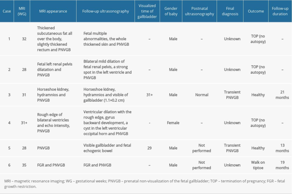
The average age of the 32 pregnant women was (31.3±4.6) years old. MRI examination was performed at (31.3±3.2) gestational weeks (GW), and the mean follow-up duration was (22.7±13.1) months. Among 32 patients, PNVGB was detected at the second trimester (n=6) and third trimester (n=26) of pregnancy. MRI examination found that 26 patients (81.3%) had isolated PNVGB (Table 1), while 6 patients (18.8%) had additional malformations (Table 2).
OUTCOMES OF 32 PATIENTS WITH PNVGB:
The outcomes of 32 patients with PNVGB are summarized in Figure 1. In 26 patients with isolated PNVGB, 7 cases were found in the gallbladder on MRI and 4 on subsequent ultrasonography. There was 1 patient with termination of pregnancy (TOP) and 1 was lost to follow-up. Of the remaining 24 patients, all had a healthy child, among whom 10 did not receive ultrasonography, 9 had normal gallbladders, 3 showed non-visualization of gallbladders, 1 had a small gallbladder, and 1 had a left small gallbladder.
Among 6 patients with additional malformations, 3 terminated their pregnancies due to combined severe malformations; 1 patient with horseshoe kidney showed a visible gallbladder on the following ultrasonography and had a healthy child with normal gallbladder; 1 patient with fetal growth restriction (FGR) did not receive ultrasonography postnatally and had a child who walked on tiptoe; 1 patient with fetal echogenic bowel was found to have a visible gallbladder on subsequent ultrasonography, but the postnatal ultrasonography was not performed, and the child was healthy.
MRI IN THE DIAGNOSIS OF PNVGB:
Among 32 patients, 7 patients (21.9%) with isolated PNVGB had visible gallbladders on MRI (Figure 2), in which 1 was lost to follow-up and the others all had a healthy child during 18–51 months of follow-up (Table 1). In 25 patients (78.1%) with non-visualized gallbladders on MRI, normal gallbladder and gallbladder agenesis found in 9 patients and 4 patients, respectively, postnatal ultrasonography was not performed in 8 patients, and TOP occurred in 4 patients, without an autopsy (due to parent refusal) (Tables 1, 2).
One patient was lost to follow-up. Twenty-seven children survived, and examination by a neonatologist revealed no jaundice, digestive tract symptoms, biliary atresia, or apparent aneuploidy. The children were healthy and well-developed at the age of 6–57 months, except for 1 child who walked on tiptoe, and this information was confirmed by telephone with the parents.
Discussion
Ultrasonography is an optimal imaging technique in prenatal diagnosis due to its non-invasiveness, no use of radiation, and real-time imaging and repeated examinations. It is usually performed several times during pregnancy. However, complicated pathological conditions or abnormalities are extremely difficult to show clearly due to the limited visual field [17]. MRI, a highly sensitive imaging method without ionizing radiation, can show the delicate fetal anatomy, such as the brain, chest, abdomen, and vasculature, and can repeatedly display obviously suspicious lesions to improve the diagnostic ability [18]. For complex fetal malformations and rare cases, MRI can provide additional information that ultrasonography is unable to show [19,20]. In the present study, 32 patients with PNVGB diagnosed by ultrasonography underwent MRI examination during the second and third trimester of pregnancy. Through MRI examination, 26 cases were found to be isolated PNVGB, among whom 7 patients presented visible gallbladders; 6 patients had additional malformations, suggesting that MRI may be valuable in identification of the fetal gallbladder.
Isolated PNVGB is reported to be related to biliary atresia in only 2 case reports [21,22]. One patient terminated the pregnancy at approximately 18 gestational weeks and the diagnosis was made based on the results of autopsy. The other had biliary atresia with an uncommon postnatal course, which might have been caused by peritonitis and ileal necrosis. In this study, 26 out of 32 cases were suggested to be isolated PNVGB, but none had biliary atresia. Among these cases, 24 patients delivered a healthy child except for 1 case of TOP and 1 patient lost to follow-up, indicating that the prognosis of patients with isolated PNVGB was favorable. Additionally, among 6 patients with additional malformations, 3 chose TOP due to combined severe abnormalities. It was thus clear that MRI can offer more information about the fetus and is superior to ultrasonography in the identification of fetal malformations.
The MRI appearance of the fetal gallbladder is alterable, and the fetal bile changes in an age-dependent manner with the signal intensity, especially after 30 GW, which may result in non-visualization of the gallbladder [16]. Additionally, there is a significant gallbladder contractility in fetuses, and the contractility cycle is about 3 hours during pregnancy [23]. It was reported that the phase of the fetal gallbladder contraction can cause non-visualization when the signal intensity of the remaining gallbladder bile is the same as that of the liver on either T2- or T1-weighted sequences [16]. In the present study, the pregnant women with visualization of gallbladder on ultrasonography underwent MRI because of some other factors, such as abnormal fetal head and fetal growth, but not PNVGB on ultrasonography; the gallbladder was found in 6 patients on subsequent prenatal ultrasonography and in 9 patients by the postnatal ultrasonography. The reason why the gallbladder was not indicated on MRI might be associated with the gallbladder contraction at each time of examination.
In this study, 5 patients with non-visualization of the gallbladder or small gallbladders through the prenatal and postnatal ultrasonography were diagnosed as having gallbladder agenesis. Among these 5 patients, a small gallbladder on MRI was visible in 1, which suggested that MRI has potential for fetal gallbladder screening. One patient with non-visualized gallbladder on both MRI and subsequent ultrasonography terminated the pregnancy at 32 gestational weeks because the risk of gallbladder atresia could not be completely ruled out, and the autopsy was not performed due to patient refusal.
This study has some limitations. First, this was a case-series study, so statistical analysis was not performed. Second, cystic fibrosis (CF) was not identified in 32 patients. CF, an autosomal recessive disease, is reported to be associated with PNVGB [13,24]. Moreover, chromosome examination was not conducted in the fetuses who were aborted, although the surviving children undergoing chromosome examination showed no signs of chromosomal abnormalities.
The findings from this retrospective study are supported by a recent systematic review of the literature [25].
Conclusions
Additional information on fetuses is crucial for prenatal diagnosis and neonatal care. MRI should be considered as an important supplementary technique when PNVGB is detected or suspected by ultrasonography.
Figures
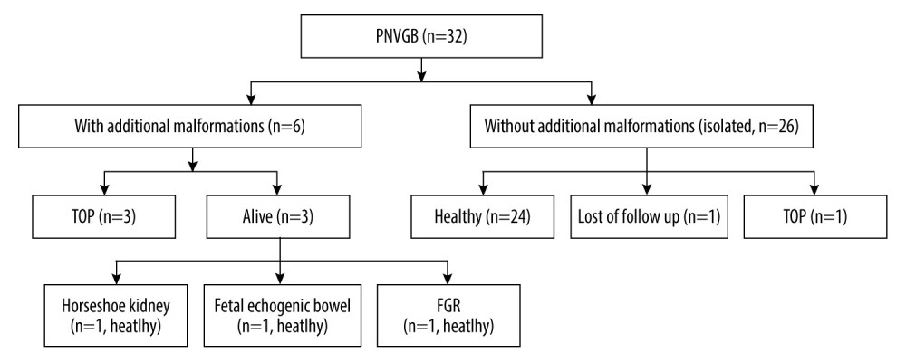 Figure 1. Outcomes of 32 cases of PNVGB. PNVGB – prenatal non-visualization of gallbladder; TOP – termination of pregnancy; FGR – fetal growth restriction.
 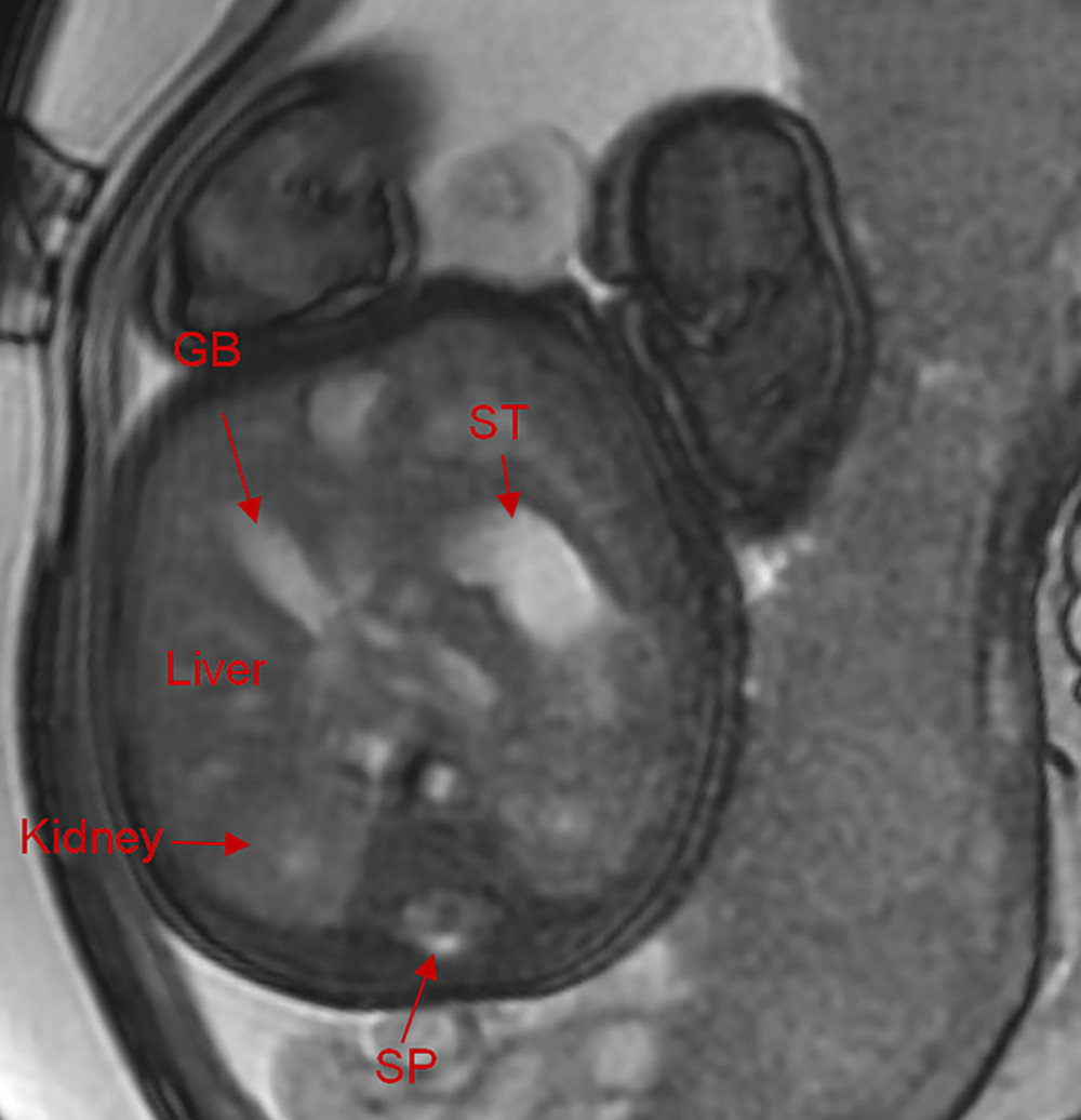 Figure 2. The MRI in 1 patient with PNVGB at 33 gestational weeks showed a normal gallbladder (3.2×1.2×1.2 cm). GB – gallbladder; ST – stomach; SP – spleen.
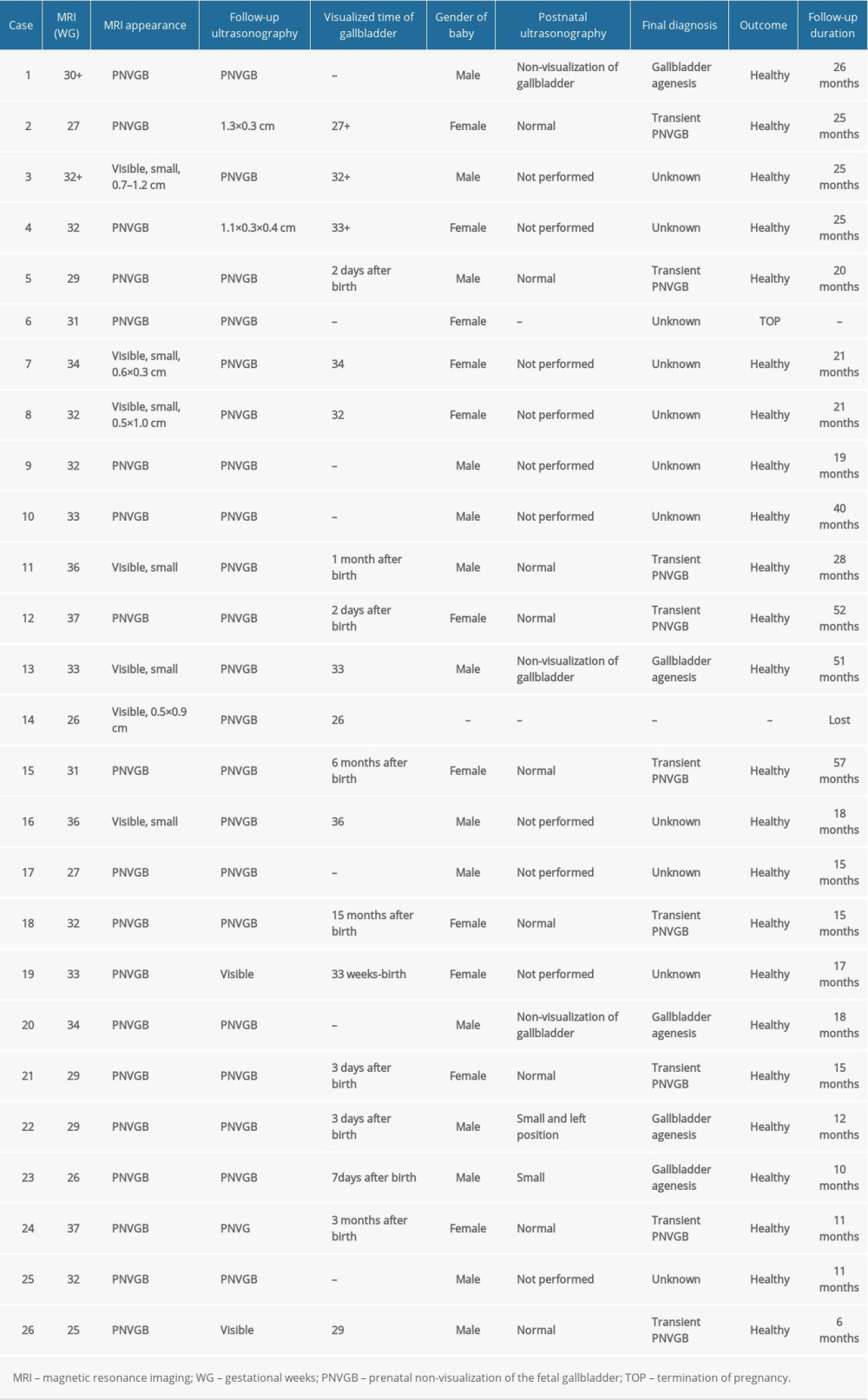
References
1. Blazer S, Zimmer EZ, Bronshtein M, Nonvisualization of the fetal gallbladder in early pregnancy: Comparison with clinical outcome: Radiology, 2002; 224(2); 379-82
2. Bardin R, Ashwal E, Davidov B, Nonvisualization of the fetal gallbladder: Can levels of gamma-glutamyl transpeptidase in amniotic fluid predict fetal prognosis?: Fetal Diagn Ther, 2016; 39(1); 50-55
3. Shen O, Rabinowitz R, Yagel S, Gal M, Absent gallbladder on fetal ultrasound: Prenatal findings and postnatal outcome: Ultrasound Obstet Gynecol, 2011; 37(6); 673-77
4. Albay S, Malas MA, Koyuncu E, Evcil EH, Morphometry of the gallbladder during the fetal period: Surg Radiol Anat, 2010; 32(4); 363-69
5. Hertzberg BS, Kliewer MA, Maynor C, McNally PJ, Nonvisualization of the fetal gallbladder: frequency and prognostic importance: Radiology, 1996; 199(3); 679-82
6. Muller F, Bernard P, Salomon LJ, Role of fetal blood sampling in cases of non-visualization of fetal gallbladder: Ultrasound Obstet Gynecol, 2015; 46(6); 743-44
7. Di Pasquo E, Kuleva M, Rousseau A, Outcome of non-visualization of fetal gallbladder on second-trimester ultrasound: Cohort study and systematic review of literature: Ultrasound Obstet Gynecol, 2019; 54(5); 582-88
8. Lakshminarayanan B, Davenport M, Biliary atresia: A comprehensive review: J Autoimmun, 2016; 73; 1-9
9. Chalouhi GE, Muller F, Dreux S, Prenatal non-visualization of fetal gallbladder: Beware of biliary atresia!: Ultrasound Obstet Gynecol, 2011; 38(2); 237-38
10. Nizery L, Chardot C, Sissaoui S, Biliary atresia: Clinical advances and perspectives: Clin Res Hepatol Gastroenterol, 2016; 40(3); 281-87
11. Chardot C, Debray D, Biliary atresia: A condition requiring urgent diagnosis and treatment: Arch Pediatr, 2011; 18(4); 476-81
12. Matar M, Ayoubi JM, Picone O, Prenatal diagnosis of gallbladder abnormalities: A review: J Gynecol Obstet Biol Reprod (Paris), 2014; 43(8); 581-86
13. Bergougnoux A, Jouannic JM, Verneau F, Isolated nonvisualization of the fetal gallbladder should be considered for the prenatal diagnosis of cystic fibrosis: Fetal Diagn Ther, 2019; 45(5); 312-16
14. Saguintaah M, Couture A, Veyrac C, MRI of the fetal gastrointestinal tract: Pediatr Radiol, 2002; 32(6); 395-404
15. Brugger PC, Prayer D, Fetal abdominal magnetic resonance imaging: Eur J Radiol, 2006; 57(2); 278-93
16. Brugger PC, Weber M, Prayer D, Magnetic resonance imaging of the fetal gallbladder and bile: Eur Radiol, 2010; 20(12); 2862-69
17. Rossi AC, Prefumo F, Accuracy of ultrasonography at 11–14 weeks of gestation for detection of fetal structural anomalies: A systematic review: Obstet Gynecol, 2013; 122(6); 1160-67
18. Griffiths PD, Bradburn M, Campbell MJ, MRI in the diagnosis of fetal developmental brain abnormalities: The MERIDIAN diagnostic accuracy study: Health Technol Assess, 2019; 23(49); 1-144
19. Valevičienė NR, Varytė G, Zakarevičienė J, Use of magnetic resonance imaging in evaluating fetal brain and abdomen malformations during pregnancy: Medicina (Kaunas), 2019; 55(2) pii: E55
20. Levine D, Barnes PD, Robertson RR, Fast MR imaging of fetal central nervous system abnormalities: Radiology, 2003; 229(1); 51-61
21. Ben-Ami M, Perlitz Y, Shalev S, Prenatal diagnosis of extrahepatic biliary duct atresia: Prenat Diagn, 2002; 22(7); 583-85
22. Boughanim M, Benachi A, Dreux S, Nonvisualization of the fetal gallbladder by second-trimester ultrasound scan: Strategy of clinical management based on four examples: Prenat Diagn, 2008; 28(1); 46-48
23. Tanaka Y, Senoh D, Hata T, Is there a human fetal gallbladder contractility during pregnancy?: Hum Reprod, 2000; 15(6); 1400-2
24. Duguépéroux I, Scotet V, Audrézet MP, Nonvisualization of fetal gallbladder increases the risk of cystic fibrosis: Prenat Diagn, 2012; 32(1); 21-28
25. Di Pasquo E, Kuleva M, Rousseau A, Outcome of non-visualization of fetal gallbladder on second-trimester ultrasound: Cohort study and systematic review of literature: Ultrasound Obstet Gynecol, 2019; 54(5); 582-88
Figures
Figure 1. Outcomes of 32 cases of PNVGB. PNVGB – prenatal non-visualization of gallbladder; TOP – termination of pregnancy; FGR – fetal growth restriction.Figure 2. The MRI in 1 patient with PNVGB at 33 gestational weeks showed a normal gallbladder (3.2×1.2×1.2 cm). GB – gallbladder; ST – stomach; SP – spleen. In Press
06 Mar 2024 : Clinical Research
Prevalence and Variation of Medical Comorbidities in Oral Surgery Patients: A Retrospective Study at Jazan ...Med Sci Monit In Press; DOI: 10.12659/MSM.943884
08 Mar 2024 : Clinical Research
Evaluation of Foot Structure in Preschool Children Based on Body MassMed Sci Monit In Press; DOI: 10.12659/MSM.943765
15 Apr 2024 : Laboratory Research
The Role of Copper-Induced M2 Macrophage Polarization in Protecting Cartilage Matrix in OsteoarthritisMed Sci Monit In Press; DOI: 10.12659/MSM.943738
07 Mar 2024 : Clinical Research
Knowledge of and Attitudes Toward Clinical Trials: A Questionnaire-Based Study of 179 Male Third- and Fourt...Med Sci Monit In Press; DOI: 10.12659/MSM.943468
Most Viewed Current Articles
17 Jan 2024 : Review article
Vaccination Guidelines for Pregnant Women: Addressing COVID-19 and the Omicron VariantDOI :10.12659/MSM.942799
Med Sci Monit 2024; 30:e942799
14 Dec 2022 : Clinical Research
Prevalence and Variability of Allergen-Specific Immunoglobulin E in Patients with Elevated Tryptase LevelsDOI :10.12659/MSM.937990
Med Sci Monit 2022; 28:e937990
16 May 2023 : Clinical Research
Electrophysiological Testing for an Auditory Processing Disorder and Reading Performance in 54 School Stude...DOI :10.12659/MSM.940387
Med Sci Monit 2023; 29:e940387
01 Jan 2022 : Editorial
Editorial: Current Status of Oral Antiviral Drug Treatments for SARS-CoV-2 Infection in Non-Hospitalized Pa...DOI :10.12659/MSM.935952
Med Sci Monit 2022; 28:e935952