22 March 2022: Clinical Research
Comparison of the Effectiveness of Endometrial Receptivity Analysis (ERA) to Guide Personalized Embryo Transfer with Conventional Frozen Embryo Transfer in 281 Chinese Women with Recurrent Implantation Failure
Yan Jia1BCDEF, Yulin Sha12BF, Zhu Qiu1BCD, Yanhua Guo1CE, Aixiang Tan1EF, Yan Huang1EF, Ying Zhong1EF, Yajun Dong1ADF, Hongxia Ye1ADG*DOI: 10.12659/MSM.935634
Med Sci Monit 2022; 28:e935634


Abstract
BACKGROUND: This study aimed to compare the effectiveness of endometrial receptivity analysis (ERA)-guided personalized embryo transfer (pET) with conventional frozen embryo transfer (FET) in 281 Chinese women with recurrent implantation failure (RIF).
MATERIAL AND METHODS: A total of 281 eligible patients with RIF were recruited and assigned to ERA (ERA followed by pET) and FET groups. The clinical pregnancy outcomes were compared between the 2 groups.
RESULTS: There were no significant differences between the ERA and FET groups in terms of endometrial thickness on the day of embryo transfer, mean attempts of assisted reproductive technology (ART) treatment, anti-Mullerian hormone, follicle-stimulating hormone, or antral follicle count in the fresh cycle (P>0.05). The ERA test identified 35% of samples as receptive and 65% as nonreceptive, and comparable pregnancy outcomes were observed between receptive and nonreceptive patients (P>0.05). Higher pregnancy and implantation rates were found in the ERA group than in the FET group (P<0.01), while no significant differences were detected between the 2 groups in terms of miscarriage rates (P>0.05).
CONCLUSIONS: In this study of Chinese women with RIF undergoing in vitro fertilization and embryo transfer, ERA-guided pET resulted in a significant improvement in pregnancy and implantation rates when compared with FET.
Keywords: Embryo Implantation, Embryo Transfer, Infertility, China, Endometrium, Female, Humans, Infertility, Female, Pregnancy, Pregnancy Rate
Background
Infertility, which is defined as a failure to conceive despite 12 months of regular, unprotected sexual intercourse, is a worldwide issue [1]. As a global public health concern, this disorder of the reproductive system is estimated to occur in 15% of all couples of reproductive age worldwide [2]. Results from the 2017 Global Burden of Disease Study showed that there were 123 085 individuals living with infertility across the globe, which was responsible for 957 000 years lived with disability [3]; more importantly, both the prevalence and the disease burden of infertility showed an upward trend from 1990 through 2017 [4]. In China, the prevalence of infertility is estimated to be 25% among couples of reproductive age, which poses heavy social, economic, family, and disease burdens in the country [5].
Although the exact cause remains unclear, multiple factors have been identified to be responsible for infertility [6], among which recurrent implantation failure (RIF) is accepted as a major cause of infertility [7–9]. Previous studies have identified many factors that contribute to the pathogenesis of RIF, and endometrial receptivity, which is crucial for embryo implantation, is widely considered as a primary cause for implantation failure [10–12]. A receptive endometrium is a prerequisite for successful embryo implantation and subsequent pregnancy [13].
The window of implantation (WOI), which is necessary for the implantation of the blastocyst in the uterus, usually starts between days 19 and 20 of the menstrual cycle, and generally lasts between 24 and 36 hours [14]. During the WOI, the endometrium expresses a sophisticated repertoire of proteins that allow it to become receptive to implantation by the embryo [15]. Precise prediction of the WOI is therefore of great significance to improve the implantation rate and the likelihood of pregnancy [16].
Endometrial receptivity analysis (ERA) was developed to identify the receptive state of the endometrium and personalize the time of embryo transfer through the profiling of the expression of 248 genes at different stages of the endometrial cycle. This assay has shown higher accuracy than histological dating in the identification of endometrial dating and receptivity [17]. More importantly, ERA results remain reproducible in the same individual across multiple menstrual cycles (from 29 through 40 months) as long as the body mass index (BMI), endometrial thickness, and treatment regimens are not changed during this period [17]. Previous studies have shown the effectiveness of ERA for the prediction of the WOI, and the ERA test was identified as an effective and reproducible approach to guiding pET [18–22]; however, the efficacy of such a tool for pET in Chinese women requires further investigation [23]. Therefore, this study aimed to compare the effectiveness of ERA-guided pET with conventional frozen embryo transfer (FET) in 281 Chinese women with RIF.
Material and Methods
ETHICS STATEMENT:
The protocol of this study has been reviewed and approved by the Ethics Review Committee of Chengdu Xi’nan Gynecology Hospital (approval no. 2020-018). Signed informed consent was obtained from all participants.
PATIENT ENROLLMENT:
Patients undergoing FET at the blastocyst stage on days 5 or 6 during the period of November 2019 through March 2021 were recruited for this study. The inclusion criteria were (1) ages of 18 through 37 years; (2) BMI between 18.5 and 30 kg/m2; (3) couples experiencing multiple embryo implantation failures (at least 2 cycles of embryo transfer, or transfer of at least 3 good-quality blastocysts with a Gardner’s score of 4BB or higher) [24]; and (4) at least 1 remaining blastocyst with a Gardner’s score of 4BB or higher. Patients with genetic disorders, anatomical abnormality of the genital tract, infections, endocrine diseases, immune disorders, or severe asthenospermia were excluded from the study. Finally, a total of 281 eligible patients were included in the study and assigned to the ERA group (n=140) or the FET group (n=141). We collected participants’ demographic features from the medical records.
ENDOMETRIAL SAMPLING AND PROCESSING:
Patients in the ERA group underwent pET, while patients in the FET group underwent conventional frozen embryo transfer. Patients in the ERA group underwent the ERA test under either a hormonal replacement therapy (HRT) cycle or a natural cycle [19]. In the HRT cycle, 50 to 70 mg of endometrial biopsy samples were collected from the uterine fundus using a sterile suction tube (Shanghai Jiaobao Medical Health Care Technology Co., Ltd.; Shanghai, China) 120 ± 3 h after the start of administration with utrogestan vaginal 300 mg capsules (CYNDEA PHARMA SL; Olvega, Spain) every 12 h and 20 mg dydrogesterone (Abbott Biologicals B.V.; Amstelveen, The Netherlands) twice a day on day 5 from the start of menstruation (P+5). In the natural cycle, endometrial specimens were sampled 7 days after the luteinizing hormone surge (LH+7) or 7 days after the administration of human chorionic gonadotropin (hCG+7). All endometrial specimens were transferred to cryotubes (Biosigma S.p.A.; Cona, Italy) containing 1.5 mL RNA later solution (Qiagen GmbH; Hilden, Germany) and were shaken vigorously to allow for the stabilization of the genetic material present in the tissue. Endometrial specimens were stored at 4°C for at least 4 h or stored at −20°C and then shipped at room temperature for the ERA test. Patients in the FET group did not receive ERA and underwent blastocyst transfer between 120 and 126 h after corpus luteum transformation.
ERA AND WOI PREDICTION:
All endometrial specimens were sent to Hangzhou Veritas Genetics Medical Institute Co., Ltd. (Hangzhou, China) for ERA. Total RNA was extracted from endometrial specimens using a QIAGEN QIA cube robotic workstation and QIAGEN spin-column kits (Qiagen; Hilden, Germany), and good-quality RNA samples (RNA integrity number R7) were employed for subsequent ERA as described previously [20].
The transcriptomic sequencing data were processed using RNASeq, and the endometrial receptivity status was assessed by the ERA computational predictor as described previously [17]. The endometrium was classified as receptive or nonreceptive according to the ERA assessment, and pET was done at the time determined by the ERA in the ERA group.
CLINICAL FOLLOW-UP AND OUTCOMES:
All patients were followed up until May 15, 2021, and the following were calculated and compared between the ERA and the FET groups: biochemical pregnancy rate, clinical pregnancy rate, implantation rate, clinical miscarriage rate, biochemical miscarriage rate, ectopic pregnancy rate, cumulative biochemical pregnancy rate, cumulative clinical pregnancy rate, cumulative implantation rate, cumulative clinical miscarriage rate, cumulative biochemical miscarriage rate, and cumulative ectopic pregnancy rate. The biochemical pregnancy rate was defined as the proportion of women who were positive for serum β-human chorionic gonadotropin (β-hCG) (>25 mIU/mL), without confirmation by vaginal ultrasound; clinical pregnancy rate was defined as the proportion of women who were positive for β-hCG, and a gestational sac was visualized by vaginal ultrasound at the fifth week of pregnancy; implantation rate was defined as the number of gestational sacs detected by vaginal ultrasound at the fifth gestational week divided by the number of embryos transferred; clinical miscarriage rate was defined as the proportion of women with spontaneous pregnancy losses in whom a gestational sac was previously observed; biochemical miscarriage rate was defined as the proportion of women with pregnancy losses in whom only the detection of β-hCG was positive, without a gestational sac visualized by vaginal ultrasound at the fifth week of pregnancy; ectopic pregnancy rate was defined as the proportion of women with pregnancies outside of the uterine cavity diagnosed by ultrasound, surgical visualization, or histopathology; cumulative biochemical pregnancy rate, cumulative clinical pregnancy rate, cumulative implantation rate, cumulative clinical miscarriage rate, cumulative biochemical miscarriage rate, and cumulative ectopic pregnancy rate were the appellate counterpart indicators (biochemical pregnancy rate, the clinical pregnancy rate, implantation rate, clinical miscarriage rate, biochemical miscarriage rate, ectopic pregnancy rate) following the same type of transfer arm into which the patient was randomized to up to a 12-month follow-up period, respectively. In addition, the blastocyst quality was assessed using Gardner’s scoring system, and a good-quality blastocyst was defined by a Gardner’s score of 4BB or higher [24].
STATISTICAL ANALYSIS:
All statistical analyses were performed using the statistical software SPSS version 21.0 (IBM Corp., Armonk, NY, USA). All measurement data were expressed as mean±standard deviations (SD), and all categorical data were described as proportions. Comparison of measurement data was done with the
Results
PATIENTS CHARACTERISTICS:
In the ERA group, patient age ranged from 24 to 37 years, BMI was between 18.55 and 29.78 kg/m2, and endometrial thickness ranged from 0.5 to 1.5 cm on the day of embryo transfer. In the FET group, patient age ranged from 23 to 37 years, BMI was between 18.73 and 29.38 kg/m2, and endometrial thickness ranged from 0.5 to 1.7 cm on the day of embryo transfer. Patients in the ERA group experienced 5.79±0.97 attempts with assisted reproductive technology (ART) treatment and patients in the FET group underwent 6.10±0.83 attempts. There were no significant differences between the ERA and FET groups in terms of age, BMI, endometrial thickness on the day of embryo transfer, mean attempts of ART treatment, or anti-Mullerian hormone (AMH), follicle-stimulating hormone (FSH), or antral follicle count (AFC) in the fresh cycle (P>0.05) (Table 1).
In addition, there were 60 patients who transferred day 6 embryos and 80 who transferred day 5 embryos in the ERA group, while 56 patients transferred day 6 embryos and 85 transferred day 5 embryos in the FET group. There were no significant differences detected in the clinical outcomes between patients who transferred day 5 embryos and patients who transferred day 6 embryos in the ERA group or in the FET group, except that a significantly higher biochemical miscarriage rate was observed in patients who transferred day 5 embryos than in those who transferred day 6 embryos in the FET group (Table 2).
ERA RESULTS AND FOLLOW-UP OUTCOMES:
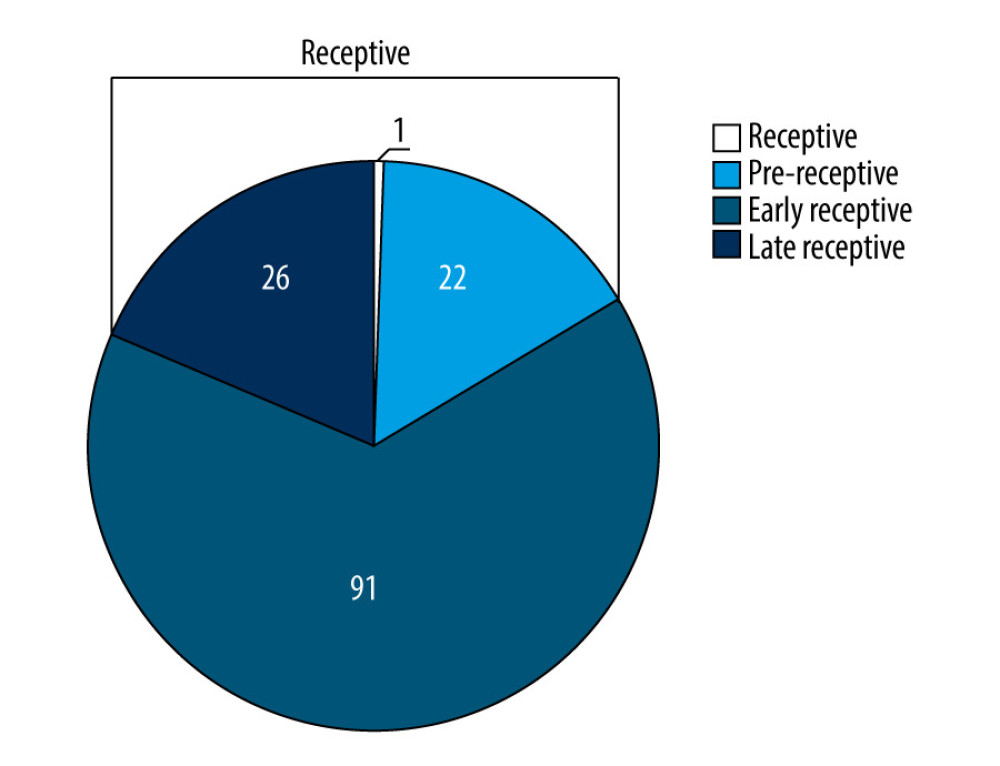
The ERA assessment presented a receptive result in 49 of the 140 patients tested (35%), and most of the nonreceptive results were pre-receptive (Figure 1). At least 1 high-quality embryo was transferred in the patients with a receptive endometrium, and the clinical follow-up showed a 48.98% clinical pregnancy rate and a 44.16% implantation rate in those patients. Among the 91 patients with a nonreceptive endometrium, the clinical follow-up showed a 50.55% clinical pregnancy rate and a 40.43% implantation rate. No ectopic pregnancies were found in patients undergoing ERA. There were no significant differences between receptive and nonreceptive patients in terms of pregnancy, implantation, or miscarriage rates (P>0.05) (Table 3).
There were no significant differences between the ERA and FET groups in terms of the number of embryos or high-quality embryos transferred per patient (P>0.05). We observed a higher pregnancy and implantation rate in the ERA group than in the FET group (P<0.01); however, no significant differences were found between the 2 groups in terms of miscarriage rate (P>0.05). In addition, no ectopic pregnancies occurred in either group (Table 4).
Then, we compared the clinical outcomes in patients in the ERA and FET groups who transferred a single embryo, and no significant differences were detected, except that a higher biochemical pregnant rate was seen in the ERA group than in the FET group (Table 5).
Discussion
In this study, our findings showed higher pregnancy and implantation rates in Chinese women with RIF who underwent ERA than in those receiving FET (
Previous studies have demonstrated that embryo- and endometrium-related factors are primary causes of RIF [8–10]. A receptive endometrium is a prerequisite for the successful implantation of embryos [13], and the endometrium is found to be most receptive during the WOI [25]. If the WOI is accurately predicted and pET is performed during the WOI, the implantation rates of transferred embryos may be improved and successful pregnancies can be achieved.
There have been numerous attempts to predict the WOI through the assessment of endometrial receptivity. Ultrasound parameters, such as endometrial thickness, volume, pattern, and vascularization, have been employed to evaluate endometrial receptivity; however, none of these parameters alone are effective to predict the WOI [26–29]. The endometrial pinopode has been proposed as a potential clinical marker to assess endometrial receptivity [30]; nevertheless, pinopodes are detectable throughout the luteal phase of the menstrual cycle [31], making them inappropriate to determine the timeframe of endometrial receptivity (WOI) [32]. Detection of serum molecules (integrin, leukemia inhibitory factor, estrogen, progesterone and their receptors, calcitonin, matrix metalloproteinase) has also been employed as an attempt to correctly identify the endometrial receptive status; however, its poor ability to predict clinical pregnancy restricts further applications of this methodology in clinical practices [33]. Additionally, even though several genes that are involved in endometrial receptivity have been identified, their diagnostic performance and value for clinical practice still remain to be elucidated [34].
Recently, a genomic tool, which consists of a customized ERA and a bioinformatic predictor for endometrial dating, was developed to evaluate endometrial receptivity through the analysis of the transcriptomic profiles of 248 selected genes. This tool has shown a global accuracy of 0.88, sensitivity of 0.90, and specificity of 0.97 [35]. Results from a comparative prospective study showed that the concordance of histological dating and ERA related to LH, used as reference, was 0.922 (range of 0.815 to 1.0), the interobserver variability between pathologists assessed by the Kappa index was 0.622 (range of 0.435 to 0.839), and the reproducibility of the ERA test was 100% consistent [17]. In a Japanese study, the pregnancy rate was 35.3% in patients with a receptive endometrium and 50% in patients with a nonreceptive endometrium after the first personalized embryo transfer was guided by the ERA test [19]. In Canada, ERA-guided pET resulted in implantation and ongoing pregnancy rates that were higher than those obtained without pET (73.7% vs 54.2% and 63.2% vs 41.7%, respectively) [13]. Results from a prospective interventional multicenter clinical trial revealed that ERA-guided pET resulted in a 51.7% pregnancy rate and a 33.9% implantation rate in RIF patients with a receptive endometrium, and a 50% pregnancy rate and a 38.5% implantation rate in those with a nonreceptive endometrium [20]. In addition, a recent 5-year multicenter open-label randomized controlled trial showed that pET guided by ERA achieved higher cumulative pregnancy rates (93.6%, 79.7%, and 80.7%,
In the present study, 281 patients with RIF were enrolled and assigned to the ERA or FET groups, and the baseline demographics and clinical characteristics were comparable between the 2 groups (
In the present study, we observed no significant differences between patients with receptive and nonreceptive endometria diagnosed by ERA in terms of pregnancy, implantation, or miscarriages (
Previous studies have shown improvements in pregnancy and implantation rates following ERA-guided pET [21,36]. A recent multicenter open-label randomized controlled trial reported significantly higher pregnancy rates at the first embryo transfer (72.5% vs 54.3%,
The present study has potential limitations. First, this was a retrospective analysis, which introduces relatively more bias than prospective clinical trials. Second, the study sample was relatively small.
Conclusions
In summary, in this study of Chinese women with RIF undergoing IVF and embryo transfer, ERA-guided pET resulted in a significant improvement in pregnancy and implantation rates when compared with FET. As a novel diagnostic tool, ERA proved to be effective in guiding pET and improving the success rates of pregnancy in patients with RIF undergoing IVF and embryo transfer.
Tables
Table 1. Comparison of the baseline patient characteristics between the endometrial receptivity analysis (ERA) and conventional frozen embryo transfer (FET) groups.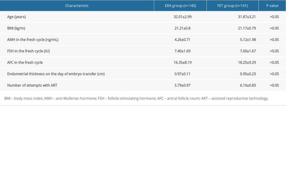 Table 2. Comparison of the clinical outcomes between transfer of day 5 and day 6 embryos in the endometrial receptivity analysis (ERA) and conventional frozen embryo transfer (FET) groups.
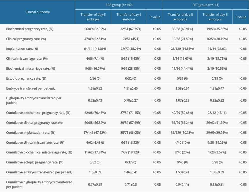 Table 3. Comparison of the clinical outcomes between the receptive and nonreceptive patients.
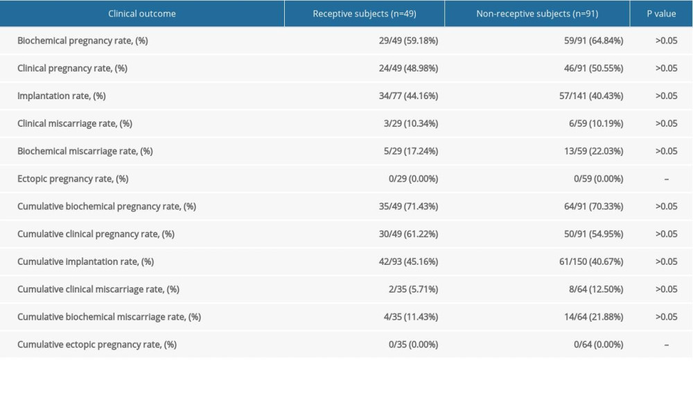 Table 4. Comparison of the clinical outcomes between the endometrial receptivity analysis (ERA) and conventional frozen embryo transfer (FET) groups.
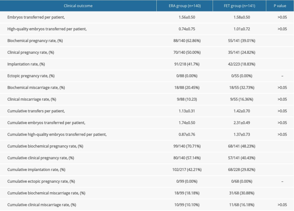 Table 5. Comparison of the clinical outcomes in patients in the endometrial receptivity analysis (ERA) and conventional frozen embryo transfer (FET) groups who transferred a single embryo.
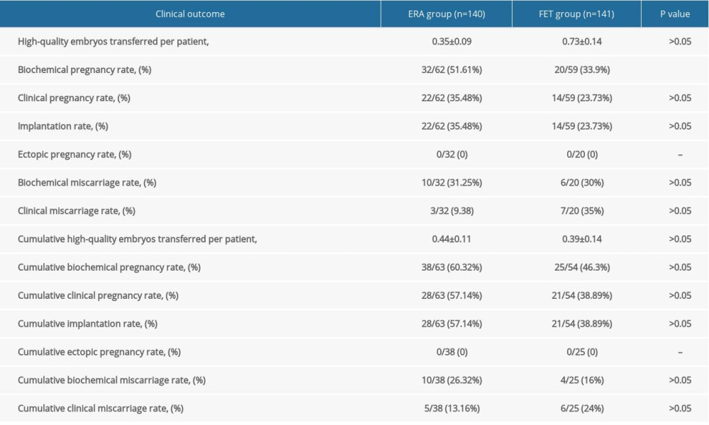
References
1. Inhorn MC, Patrizio P: Hum Reprod Update, 2015; 21; 411-26
2. Vander BM, Wyns C, Fertility and infertility: Definition and epidemiology: Clin Biochem, 2018; 62; 2-10
3. GBD 2017 Disease and Injury Incidence and Prevalence Collaborators, Global, regional, and national incidence, prevalence, and years lived with disability for 354 diseases and injuries for 195 countries and territories, 1990–2017: A systematic analysis for the Global Burden of Disease Study 2017: Lancet, 2018; 392; 1789-858
4. Sun H, Gong TT, Jiang YT, Global, regional, and national prevalence, and disability-adjusted life-years for infertility in 195 countries and territories, 1990–2017: Results from a global burden of disease study, 2017: Aging (Albany NY), 2019; 11; 10952-91
5. Zhou Z, Zheng D, Wu H, Epidemiology of infertility in China: A population-based study: BJOG, 2018; 125; 432-41
6. Tamrakar SR, Bastakoti R, Determinants of infertility in couples: J Nepal Health Res Counc, 2019; 17; 85-89
7. Coughlan C, Ledger W, Wang Q, Recurrent implantation failure: Definition and management: Reprod Biomed Online, 2014; 28; 14-38
8. Bashiri A, Halper KI, Orvieto R, Recurrent implantation failure-update overview on etiology, diagnosis, treatment, and future directions: Reprod Biol Endocrinol, 2018; 16; 121
9. Laufer N, Simon A, Recurrent implantation failure: Current update and clinical approach to an ongoing challenge: Fertil Steril, 2012; 97; 1019-20
10. Timeva T, Shterev A, Kyurkchiev S, Recurrent implantation failure: The role of the endometrium: J Reprod Infertil, 2014; 15; 173-83
11. Bellver J, Simón C, Implantation failure of endometrial origin: What is new?: Curr Opin Obstet Gynecol, 2018; 30; 229-36
12. Kliman HJ, Frankfurter D, Clinical approach to recurrent implantation failure: Evidence-based evaluation of the endometrium: Fertil Steril, 2019; 111; 618-28
13. Teh WT, McBain J, Rogers P, What is the contribution of embryo-endometrial asynchrony to implantation failure?: J Assist Reprod Genet, 2016; 33; 1419-30
14. Díaz-Gimeno P, Ruiz-Alonso M, Sebastian-Leon P, Window of implantation transcriptomic stratification reveals different endometrial subsignatures associated with live birth and biochemical pregnancy: Fertil Steril, 2017; 108; 703-10e3
15. Lessey BA, Endometrial receptivity and the window of implantation: Best Pract Res Clin Obstet Gynaecol, 2000; 14; 775-88
16. Devyatova EA, Tsaturova KA, Vartanyan EV, Predicting of successful implantation at IVF cycles: Gynecol Endocrinol, 2016; 32; 27-29
17. Díaz-Gimeno P, Horcajadas JA, Martínez-Conejero JA, A genomic diagnostic tool for human endometrial receptivity based on the transcriptomic signature: Fertil Steril, 2011; 95; 50-60
18. Díaz-Gimeno P, Ruiz-Alonso M, Blesa D, The accuracy and reproducibility of the endometrial receptivity array is superior to histology as a diagnostic method for endometrial receptivity: Fertil Steril, 2013; 99; 508-17
19. Hashimoto T, Koizumi M, Doshida M, Efficacy of the endometrial receptivity array for repeated implantation failure in Japan: A retrospective, two-centers study: Reprod Med Biol, 2017; 16; 290-96
20. Ruiz-Alonso M, Blesa D, Díaz-Gimeno P, The endometrial receptivity array for diagnosis and personalized embryo transfer as a treatment for patients with repeated implantation failure: Fertil Steril, 2013; 100; 818-24
21. Simón C, Gómez C, Cabanillas S, A 5-year multicentre randomized controlled trial comparing personalized, frozen and fresh blastocyst transfer in IVF: Reprod Biomed Online, 2020; 41; 402-15
22. Ruiz-Alonso M, Valbuena D, Gomez C, Endometrial receptivity analysis (ERA): Data versus opinions: Hum Reprod Open, 2021; 2021; hoab011
23. Jia Y, Sha YL, Qiu Z, Endometrial receptivity analysis for personalized embryo transfer in patients with recurrent implantation failure: A retrospective analysis of a Chinese cohort: Human Reprod, 2021; 36; deab130.312
24. Gardner DK, Lane M, Stevens J, Blastocyst score affects implantation and pregnancy outcome: Towards a single blastocyst transfer: Fertil Steril, 2000; 73; 1155-58
25. Rincon A, Clemente-Ciscar M, Gomez E, What is the real length of the window of implantation (WOI) in humans? Human Reproduction: England: Oxford Univ Press, 2018; 33; 360
26. Järvelä IY, Sladkevicius P, Kelly S, Evaluation of endometrial receptivity during in-vitro fertilization using three-dimensional power Doppler ultrasound: Ultrasound Obstet Gynecol, 2005; 26; 765-69
27. Kupesic S, Bekavac I, Bjelos D, Assessment of endometrial receptivity by transvaginal color Doppler and three-dimensional power Doppler ultrasonography in patients undergoing in vitro fertilization procedures: J Ultrasound Med, 2001; 20; 125-34
28. Ng EH, Chan CC, Tang OS, The role of endometrial and subendometrial vascularity measured by three-dimensional power Doppler ultrasound in the prediction of pregnancy during frozen-thawed embryo transfer cycles: Hum Reprod, 2006; 21; 1612-17
29. Mercé LT, Barco MJ, Bau S, Are endometrial parameters by three-dimensional ultrasound and power Doppler angiography related to in vitro fertilization/embryo transfer outcome?: Fertil Steril, 2008; 89; 111-17
30. Nikas G, Endometrial receptivity: Changes in cell-surface morphology: Semin Reprod Med, 2000; 18; 229-35
31. Quinn C, Ryan E, Claessens EA, The presence of pinopodes in the human endometrium does not delineate the implantation window: Fertil Steril, 2007; 87; 1015-21
32. Quinn CE, Casper RF, Pinopodes: A questionable role in endometrial receptivity: Hum Reprod Update, 2009; 15; 229-36
33. Craciunas L, Gallos I, Chu J, Coomarasamy A, Conventional and modern markers of endometrial receptivity: A systematic review and meta-analysis: Hum Reprod Update, 2019; 25; 202-23
34. Altmäe S, Koel M, Võsa U, Meta-signature of human endometrial receptivity: A meta-analysis and validation study of transcriptomic biomarkers: Sci Rep, 2017; 7; 10077
35. Clemente-Ciscar M, Ruiz-Alonso M, Blesa D, Endometrial receptivity analysis (ERA) using a next generation sequencing (NGS) predictor improves reproductive outcome in recurrent implantation failure (RIF) patients when compared to ERA arrays: Hum Reprod, 2018; 33; 8
36. Tan J, Kan A, Hitkari J, The role of the endometrial receptivity array (ERA) in patients who have failed euploid embryo transfers: J Assist Reprod Genet, 2018; 35; 683-92
37. Patel JA, Patel AJ, Banker JM, Personalized embryo transfer helps in improving in vitro fertilization/ICSI outcomes in patients with recurrent implantation failure: J Hum Reprod Sci, 2019; 12; 59-66
38. Cohen AM, Ye XY, Colgan TJ, Comparing endometrial receptivity array to histologic dating of the endometrium in women with a history of implantation failure: Syst Biol Reprod Med, 2020; 66; 347-54
39. Li HH, Liu S, Li Y, Research progress of factors affecting the endometrial receptivity: Reprod Controcep, 2016; 36; 833-38
40. Kuroda K, Horikawa T, Moriyama A, Impact of chronic endometritis on endometrial receptivity analysis results and pregnancy outcomes: Immun Inflamm Dis, 2020; 8; 650-58
41. Stankewicz T, Adaniya G, Cinnioglu C, Intramuscular versus vaginal progesterone: Can we expect differences in endometrial receptivity?: Fertil Steril, 2019; 111; E28
42. Riestenberg C, Kroener L, Quinn M, Routine endometrial receptivity array in first embryo transfer cycles does not improve live birth rate: Fertil Steril, 2021; 115; 1001-6
Tables
Table 1. Comparison of the baseline patient characteristics between the endometrial receptivity analysis (ERA) and conventional frozen embryo transfer (FET) groups.Table 2. Comparison of the clinical outcomes between transfer of day 5 and day 6 embryos in the endometrial receptivity analysis (ERA) and conventional frozen embryo transfer (FET) groups.Table 3. Comparison of the clinical outcomes between the receptive and nonreceptive patients.Table 4. Comparison of the clinical outcomes between the endometrial receptivity analysis (ERA) and conventional frozen embryo transfer (FET) groups.Table 5. Comparison of the clinical outcomes in patients in the endometrial receptivity analysis (ERA) and conventional frozen embryo transfer (FET) groups who transferred a single embryo.Table 1. Comparison of the baseline patient characteristics between the endometrial receptivity analysis (ERA) and conventional frozen embryo transfer (FET) groups.Table 2. Comparison of the clinical outcomes between transfer of day 5 and day 6 embryos in the endometrial receptivity analysis (ERA) and conventional frozen embryo transfer (FET) groups.Table 3. Comparison of the clinical outcomes between the receptive and nonreceptive patients.Table 4. Comparison of the clinical outcomes between the endometrial receptivity analysis (ERA) and conventional frozen embryo transfer (FET) groups.Table 5. Comparison of the clinical outcomes in patients in the endometrial receptivity analysis (ERA) and conventional frozen embryo transfer (FET) groups who transferred a single embryo. In Press
05 Mar 2024 : Clinical Research
Role of Critical Shoulder Angle in Degenerative Type Rotator Cuff Tears: A Turkish Cohort StudyMed Sci Monit In Press; DOI: 10.12659/MSM.943703
06 Mar 2024 : Clinical Research
Comparison of Outcomes between Single-Level and Double-Level Corpectomy in Thoracolumbar Reconstruction: A ...Med Sci Monit In Press; DOI: 10.12659/MSM.943797
21 Mar 2024 : Meta-Analysis
Economic Evaluation of COVID-19 Screening Tests and Surveillance Strategies in Low-Income, Middle-Income, a...Med Sci Monit In Press; DOI: 10.12659/MSM.943863
10 Apr 2024 : Clinical Research
Predicting Acute Cardiovascular Complications in COVID-19: Insights from a Specialized Cardiac Referral Dep...Med Sci Monit In Press; DOI: 10.12659/MSM.942612
Most Viewed Current Articles
17 Jan 2024 : Review article
Vaccination Guidelines for Pregnant Women: Addressing COVID-19 and the Omicron VariantDOI :10.12659/MSM.942799
Med Sci Monit 2024; 30:e942799
14 Dec 2022 : Clinical Research
Prevalence and Variability of Allergen-Specific Immunoglobulin E in Patients with Elevated Tryptase LevelsDOI :10.12659/MSM.937990
Med Sci Monit 2022; 28:e937990
16 May 2023 : Clinical Research
Electrophysiological Testing for an Auditory Processing Disorder and Reading Performance in 54 School Stude...DOI :10.12659/MSM.940387
Med Sci Monit 2023; 29:e940387
01 Jan 2022 : Editorial
Editorial: Current Status of Oral Antiviral Drug Treatments for SARS-CoV-2 Infection in Non-Hospitalized Pa...DOI :10.12659/MSM.935952
Med Sci Monit 2022; 28:e935952