08 January 2022: Clinical Research
Oblique Lateral Interbody Fusion with Anterolateral Screw Fixation Is as Effective as with Posterior Percutaneous Pedicle Screw Fixation in Treating Single-Segment Mild Degenerative Lumbar Diseases
Yunshan Guo 1CG* , Xiaodong Wang 1BF* , Yibing Li 1CE , Kuo Jiang 1DE , Bo Chen 2D , Jing An 2CF , Dingjun Hao 1AG* , Huimin Hu 1AE*DOI: 10.12659/MSM.934985
Med Sci Monit 2022; 28:e934985
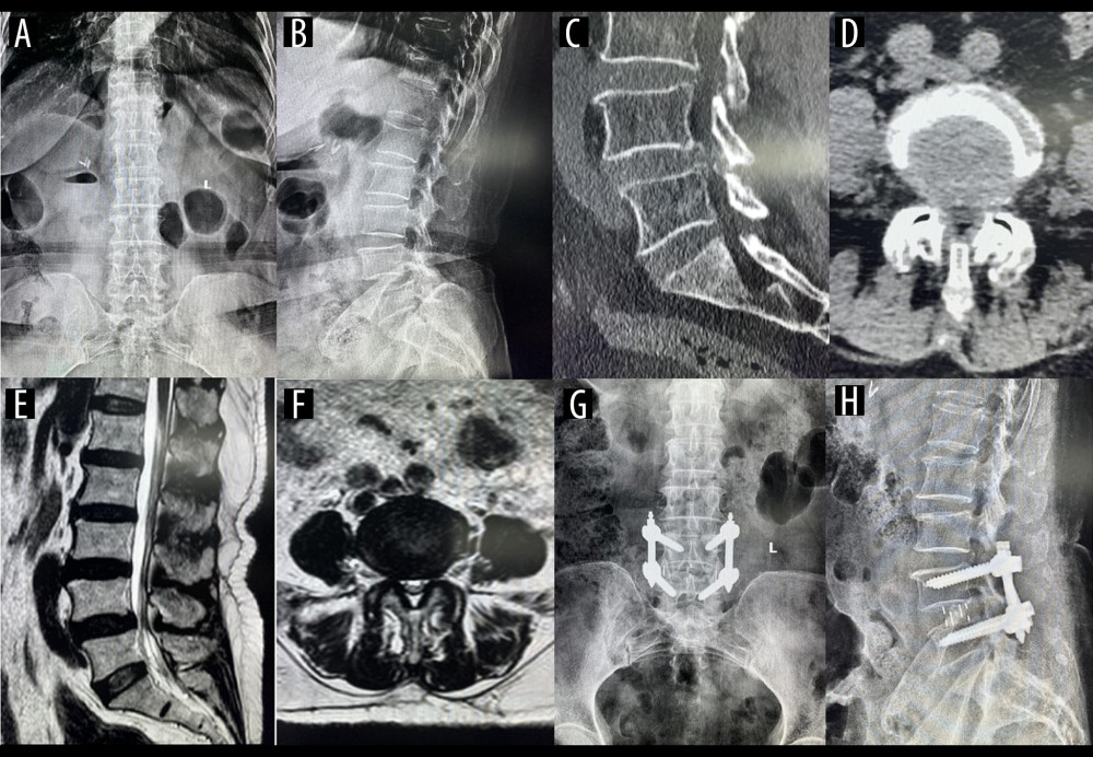
Figure 6 A typical case of oblique lateral interbody fusion with percutaneous pedicle screw fixation (OLIF+PF)A 57-year-old man had low back pain for 3 years, aggravated for 5 months. OLIF+PF was employed to treat lumbar spondylolisthesis at L4/5. (A) Preoperative anteroposterior radiograph. (B) Preoperative lateral radiograph shows L4 vertebral body I° anterior spondylolisthesis and loss of intervertebral disc height at L4/5. Before surgery, the anterior disc height (ADH) was 5.7 mm, the posterior disc height (PDH) was 5.1 mm, and the foraminal height (FH) was 13.9 mm. (C) Preoperative sagittal computed tomography (CT). (D) Preoperative cross-section CT shows disc herniation, obvious proliferation of the ligamentum flavum, and spinal canal stenosis at L4/5. (E) Preoperative sagittal magnetic resonance imaging (MRI). (F) Preoperative cross-section MRI shows disc herniation, obvious proliferation of the ligamentum flavum, and spinal canal stenosis at L4/5. (G) Postoperative anteroposterior radiograph. (H) Postoperative lateral radiograph shows the intervertebral cage at L4/5 was well in place, and the ADH, PDH, and FH were significantly increased. After surgery, the ADH was 11.2 mm, the PDH was 10.5 mm, and the FH was 19.1 mm.