17 October 2021: Clinical Research
White Matter Abnormalities in Traumatic Subarachnoid Hemorrhage: A Tract-Based Spatial Statistics Study
Min Son Kim 1ABCDE , Min Jye Cho 1CDE* , Jae Woon Kim 2CDF , Sung Ho Jang 1AEF**DOI: 10.12659/MSM.933959
Med Sci Monit 2021; 27:e933959
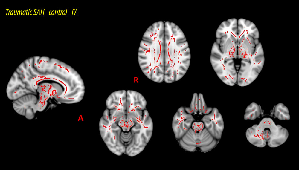
Figure 1 Results of tract-based spatial statics analysis comparing fractional anisotropy (FA) values between the patient and control groups. FA values are acquired for 48 regions of interest (ROIs) applying the white matter atlases standard template of Johns Hopkins University through the Functional Magnetic Resonance Imaging of the Brain Software Library (FSL version 5.1). The red voxels represent areas where the mean FA values are significantly higher in the control group than in the patient group. The FA values of 19 of 48 ROIs (splenium, genu, and body of the corpus callosum, both posterior limbs of the internal capsule, both corticospinal tracts, both superior corona radiata, both superior cerebellar peduncles, the middle cerebellar peduncle, the left anterior limb of the internal capsule, the left external capsule, the right cingulum, the left superior longitudinal fasciculus, the left cingulum, the left cerebral peduncle, the right posterior corona radiata) in the control group are higher than those of the patient group.