20 October 2021: Clinical Research
Analysis of Characteristics of Patients with Non-ST-Segment Elevation Myocardial Infarction by Cardiac Magnetic Resonance Imaging
Shujuan Dong 1ACE* , Yunbo Liu 2E* , Wenjing Sun 1BE* , Chunqiu Wang 3BD* , Yan Wang 3C , Wenbo Zhao 1F , Shenghui Zhao 1F , Yingjie Chu 1G*DOI: 10.12659/MSM.933220
Med Sci Monit 2021; 27:e933220
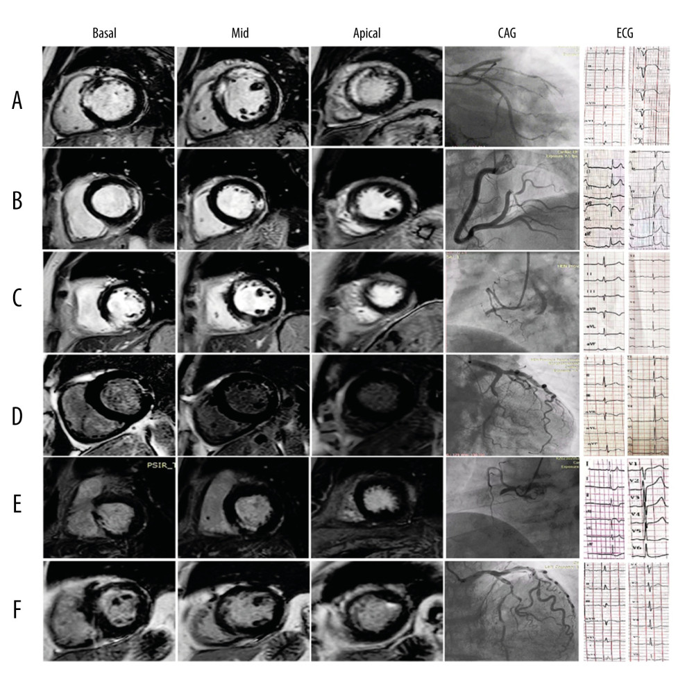
Figure 2 Images from 6 AMI patients. (A) ECG in leads V1–V5 manifests ST-elevation in a 57-year-old man. CMR illustrates transmural infarction in the anterior segments detected by DE-MRI. Coronary angiography revealed LAD 100% occluded proximally. (B) ST-segment elevation on ECG in leads II, III, and aVF in a 48-year-old man. CMR illustrates contrast-enhanced areas in the lateral segments characterized by DE-MRI. Coronary angiography revealed a 99% distal stenosis in the RCA. (C) A 58-year-old MI patient with a normal ST segment. CMR shows contrast-enhanced areas in the inferior segments characterized by DE-MRI. Coronary angiography revealed 100% proximal occluded RCA. (D) A 55-year-old woman with MI with a normal ST segment. CMR illustrates non-transmural hyperenhancement in the lateral segment. Coronary angiography revealed 95% middle stenosis in the LCx. (E) ECG of a 50-year-old patient showing presence ST-depression in inferior leads. CMR showed partial transmural and subendocardial necrosis in the inferior segments detected by DE-MRI. Coronary angiography revealed 100% proximal occluded RCA. (F) ECG of a 66-year-old woman presenting non-ST-segment elevation in anterior leads. CMR showed non-transmural hyperenhancement in the anterior segment detected by DE-MRI. Coronary angiography revealed 95% proximal stenosis in the LAD. DE-MRI – delayed-enhancement magnetic resonance imaging; MI – myocardial infarction; LAD – left anterior descending artery; LCx – left circumflex; RCA – right coronary artery.