22 October 2021: Database Analysis
Comparison of Magnetic Resonance Imaging of the Lower Uterine Segment in Pregnant Women with Central Placenta Previa with and without Placenta Accreta Spectrum from a Single Center
Shunyu Hou 1G* , Ye Song 1B* , Jiahui Wu 2D , Liping Zhou 1B , Suya Kang 1F , Xi Chen 3C , Lei Zhang 4F , Yanli Lu 5C , Yongfei Yue 1ACE*DOI: 10.12659/MSM.932759
Med Sci Monit 2021; 27:e932759
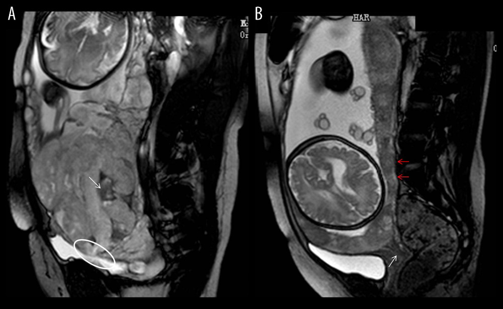
Figure 4 Magnetic resonance imaging (MRI) characteristics in central placenta previa patients with and without placenta accreta (Sagittal T2-weighted). (Microsoft Office PowerPoint 2007, Microsoft, USA). (A) Central placenta previa with placenta accrete: Placenta thickness is 12.3 centimeters; The cervical canal is almost completely eroded by the placenta and we can hardly see a clear cervix; The placental signal is heterogeneous and many dark T2 bands are seen in the placenta (white arrow); The whole placenta is located in the lower part of the uterus, which is dilated; A partial myometrium defect can be seen at the uterine and placental interface (white circle); (B) Central placenta previa without placenta accrete: Placenta thickness is 3.2 centimeters; We can see a clear cervix (white arrow); Placenta signals are homogeneous; The boundary between the uterine myometrium and placenta is clear (red arrow).