24 September 2021: Clinical Research
Comparison of Outcomes Following TiRobot-Assisted Sacroiliac Screw Fixation with Bone Grafting and Traditional Screw Fixation without Bone Grafting for Unstable Osteoporotic Sacral Fracture: A Single-Center Retrospective Study of 33 Patients
Zhaojie Liu ABE* , Ya Gu CDF* , Xin Jin DF , Wei Tian BE , Haotian Qi CD , Yuxi Sun BF , Gang Li DF , Hongchuan Wang BF , Xiang Xiao DF , Pengfei Li EF , Yongcheng Hu AD , Jian Jia AE*DOI: 10.12659/MSM.932724
Med Sci Monit 2021; 27:e932724
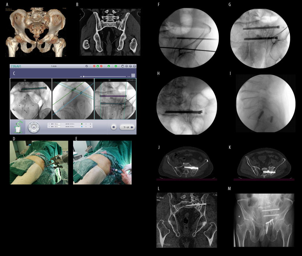
Figure 4 A 75-year-old man with an injury from a car accident. (A) Computed tomography (CT) construction scan of the pelvic ring reveals the displaced pubic body and inferior pubic rami fracture associated with the ipsilateral sacral fracture. (B) Coronal CT scan showing the complete sacral fracture. (C) Intraoperative planning of sacroiliac screws after collection and transmission of the radiographs. (D) The view of robotic arm positioning at the entry point of sacroiliac screw. (E) The intraoperative picture demonstrating bone grafting injected along the channel of the sacroiliac screw. (F) Bone grafting along sacroiliac screw channel under C-arm fluoroscopy. (G–I) Intraoperative anteroposterior, inlet, and outlet views. (J–L) Postoperative coronal and transverse CT views revealing the satisfactory positioning of sacroiliac screws and the bone grafting. (M) The pelvic outlet view at 6 months after surgery showing that the fracture healed.