24 September 2021: Clinical Research
Comparison of Outcomes Following TiRobot-Assisted Sacroiliac Screw Fixation with Bone Grafting and Traditional Screw Fixation without Bone Grafting for Unstable Osteoporotic Sacral Fracture: A Single-Center Retrospective Study of 33 Patients
Zhaojie Liu ABE* , Ya Gu CDF* , Xin Jin DF , Wei Tian BE , Haotian Qi CD , Yuxi Sun BF , Gang Li DF , Hongchuan Wang BF , Xiang Xiao DF , Pengfei Li EF , Yongcheng Hu AD , Jian Jia AE*DOI: 10.12659/MSM.932724
Med Sci Monit 2021; 27:e932724
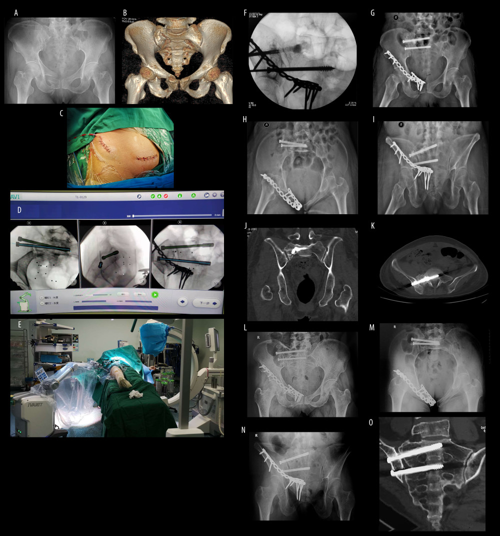
Figure 3 A 67-year-old woman with injury from falling from a height. (A) The anteroposterior pelvic view reveals displaced superior and inferior pubic rami fractures on the right side, close to pubic symphysis. (B) Computed tomography (CT) construction scan of the pelvic ring demonstrates the overlapping superior and inferior pubic rami fracture and the compression sacral fracture due to the internal rotation of the pelvis. It concerns a type B1 lesion according to Tile classification on pelvic fracture. (C) The medial and lateral incisions of anterior pelvis ring. (D) Path planning of sacroiliac screw placement in S1 and S2. (E) Overall view of robot-aided sacroiliac screw manipulation. (F) Bone grafting via sacroiliac screw channel under C-arm fluoroscopy. There is a dense shadow in sacral ala, showing the injectable bone substitute. (G–I) Postoperative anteroposterior, inlet and outlet views. (J, K) Coronal and transverse CT views showing the positioning of the sacroiliac screw and the bone grafting. (L–O) Anteroposterior, inlet and outlet views and CT scan at 6 months after surgery showing the union of sacrum.