13 October 2021: Clinical Research
Comparing the Traditional Versus Conservative Endodontic Access Cavities Design of the Maxillary First Molar: Using Cone-Beam Computed Tomography
Huachao Sui 1ABE* , Bo Zhao 2ABE* , Haidan Nie 1BE* , Xin Hao 1BG , Feng Qiao 3C , Cuicui Sun 1C , Changyi Li 4G , Liwen Zhou 1E , Ligeng Wu 1AG*DOI: 10.12659/MSM.932410
Med Sci Monit 2021; 27:e932410
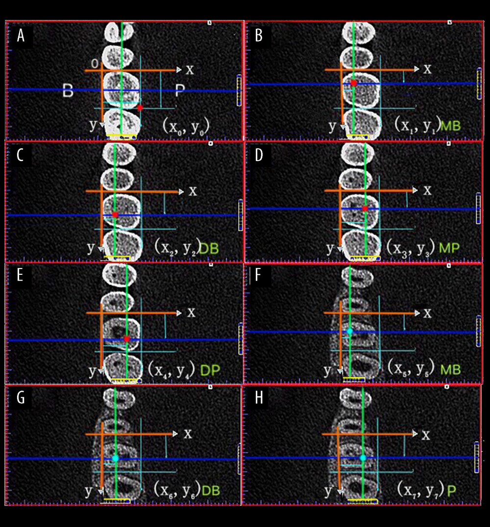
Figure 2 Each pulp horn and root canal orifice was identified according to the first appearance of their low-density shadow and was designated by the x, y coordinates in the X and Y axes system. (A) Buccal-lingual distance; Mesial-distal distance. (B–E) Pulp horns (red dots): the intersection of the coronal plane (blue line) and the sagittal plane (green line) (MB, DB, MP, DB). (F–H) Root canal orifices (blue dots): the intersection of the coronal plane (blue line) and the sagittal plane (green line) (MB, DB, P). MB – mesiobuccal; DB – distobuccal; MP – mesiopalatal; DP – distopalatal; P – palatal.