01 October 2021: Clinical Research
Comparison of the Use of Magnetic Resonance Imaging of Partial Anterior Cruciate Ligament Tears Using Maximum Knee Flexion in the Lateral Decubitus Position with Routine Knee Positioning
Zijun Xu 1ABCDEFG* , Yichao Chen 2ABCDEF* , Jianghua Zhu 3FG , Lin Zhang 1FG* , Peng Wu 2AG*DOI: 10.12659/MSM.932228
Med Sci Monit 2021; 27:e932228
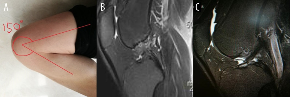
Figure 1 (A) The sketch of maximum flexion of knee joint with maximum flexibility of 150°. (B) Image from a 37-year-old man, 13 months after injury. Only routine position magnetic resonance imaging (MRI) scans were provided. The display angle of anterior cruciate ligament attachment points was poor. Sagittal and coronal scanning images both had effusion disturbing the diagnosis. (C) Image from a 45-year-old man. Only routine position MRI scans were provided. The joint effusion or hemorrhage near the ligament tear site interfered with the diagnosis.