24 June 2021: Clinical Research
An Individualized Contrast-Enhanced Liver Computed Tomography Imaging Protocol Based on Body Mass Index in 126 Patients Seen for Liver Cirrhosis
Jian Jiang ABCEG , Maowei Zhang BD , Yuan Ji BCDF , Chunfeng Li CD , Xin Fang BF , Shuyuan Zhang B , Wei Wang CF , Lijun Wang EF , Ailian Liu ACEF*DOI: 10.12659/MSM.932109
Med Sci Monit 2021; 27:e932109
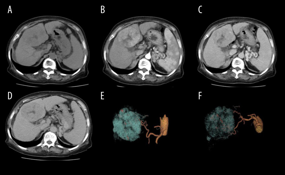
Figure 4 Scans from a 75-year-old woman (body mass index, 28.1 kg/m2) with hepatitis B cirrhosis. Liver CT was performed at 120 kV (size-specific dose estimate, 16.04 mGy) and contrast medium 550 mg I/kg. (A) Non-enhanced, (B) late arterial, (C) portal venous, and (D) delay phase images; and (E, F) 3D volume-rendering of reconstruction images, with arteries in yellow and tumor in blue. A hypervascular tumor can be seen in the liver, with hypodensity (A), hypervascularity (B), and washout (C, D) relative to the liver parenchyma. Three-dimensional volume-rendering reconstruction images show the relationship between the arteries and the tumor (E, F). The tumor diagnosis of hepatocellular carcinoma was confirmed by pathology.