28 August 2021: Clinical Research
Characteristics of the Computed Tomography Imaging Findings in 72 Patients with Airway-Invasive Pulmonary Aspergillosis
Jing Wu 1ABCDEF , Tao Zhang 1BCDEF , Junping Pan 2ABCD , Qian Zhang 3B , Xin Lin 4B , Ligong Chang 5B , Yu-Chen Chen 1ACDF* , Xinying Xue 6ADF*DOI: 10.12659/MSM.931162
Med Sci Monit 2021; 27:e931162
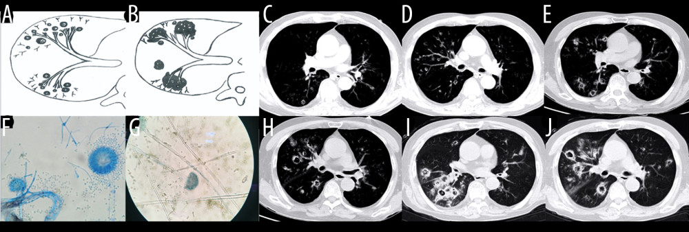
Figure 6 Case 5. An 83-year-old man had chronic obstructive pulmonary disease for more than 20 years had been aggravated for 10 days, with fever of 38°C and shortness of breath. (A, B) Schematic diagram of type III airway-invasive pulmonary aspergillosis, which showed early stage of type III and its progress. (C, D) Computed tomography (CT) scan images on the second day of admission, showing diffuse acinar nodules with punctate cavities in peripheral lungs, with internal separation. (E, H) CT scan images taken on day 5 after admission and reexamination after anti-infective treatment, showing the lung window of the same slice, specifically the lesion and the cavity were further enlarged with compartmentalized changes in the cavity. (I, J) CT scan images taken on the day 8 after admission showing the lung window on the same slice; the lesion was further enlarged, with fusion of small lesions, and the cavity was enlarged and increased, with obvious internal separation. (F, G) Sputum specimens, conidia of Aspergillus flavus; Lactophenol cotton blue dyeing, ×1000.