28 August 2021: Clinical Research
Characteristics of the Computed Tomography Imaging Findings in 72 Patients with Airway-Invasive Pulmonary Aspergillosis
Jing Wu 1ABCDEF , Tao Zhang 1BCDEF , Junping Pan 2ABCD , Qian Zhang 3B , Xin Lin 4B , Ligong Chang 5B , Yu-Chen Chen 1ACDF* , Xinying Xue 6ADF*DOI: 10.12659/MSM.931162
Med Sci Monit 2021; 27:e931162
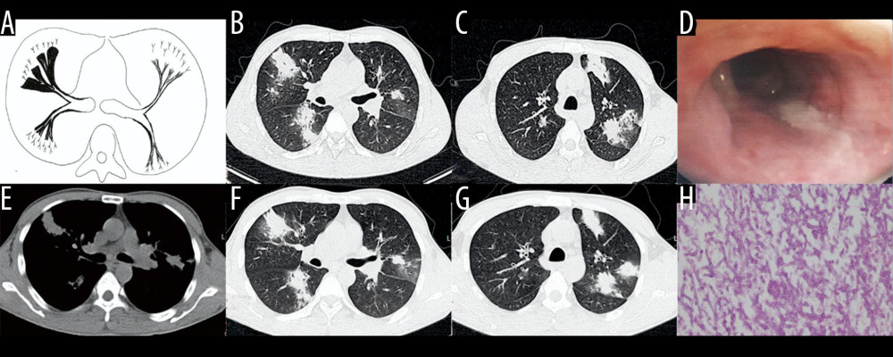
Figure 5 Case 4. A 45-year-old man had cough, sputum, and fever for 2 weeks, accompanied by shortness of breath, and he had been previously in good health. (A) Schematic diagram of type IIb airway-invasive pulmonary aspergillosis. (B, C) Computed tomography (CT) scan images taken at admission, showing both lungs with diffuse lesions distributed along the long axis of the bronchial alignment, with consolidation opacity and stenosis of the bronchus. (B) Arrow shows the location of the pathology. (D) The bronchial wall in the segmental bronchus was covered by pus-like exudates via bronchoscopy. (E) Mediastinal window shows the lesion distributed along the bronchus, and the edge was straight, consistent with the inflammatory changes. (F, G) CT scan images of the lung window on the same slice after 5 days of anti-infective treatment, showing no obvious improvement, diffuse lesions that are distributed along the periphery of the bronchus, and no bronchiectasis. (H) pathological Aspergillus hyphae and small round spores (HE ×400).