28 August 2021: Clinical Research
Characteristics of the Computed Tomography Imaging Findings in 72 Patients with Airway-Invasive Pulmonary Aspergillosis
Jing Wu 1ABCDEF , Tao Zhang 1BCDEF , Junping Pan 2ABCD , Qian Zhang 3B , Xin Lin 4B , Ligong Chang 5B , Yu-Chen Chen 1ACDF* , Xinying Xue 6ADF*DOI: 10.12659/MSM.931162
Med Sci Monit 2021; 27:e931162
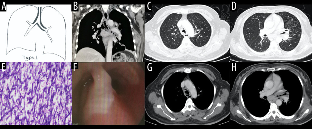
Figure 2 Case 1. A 48-year-old woman had cough and shortness of breath for 1 month, with a past history of diabetes mellitus. (A, B) The coronal view of the mediastinum, indicating major thickening along the left main bronchus, partial stenosis of the lumen, and inflammation of the distal lung tissue, which is similar to the schematic diagram. (C, D) Thickening at the walls of the trachea and left main bronchus in the lung window. (E) Pathological Aspergillus hyphae and small round spores (HE ×400). (F) The trachea and left main bronchus were covered by pus-like exudates via bronchoscopy. (G, H) Thickening of the wall of the bronchus, the soft tissue shadow in the mediastinum (inflammatory infiltration or lymph node enlargement) and extraluminal air, indicating the formation of mediastinitis and bronchial fistula.