02 May 2021: Clinical Research
Parameters Indicating Development of Influenza-Associated Acute Necrotizing Encephalopathy: Experiences from a Single Center
Suyun Li 1AE* , Dandan Hu 2AE* , Peiqing Li 1AE* , Weiqiang Xiao 3AE* , Huixian Li 4ACE* , Guangming Liu 1C , Yongling Song 1C , Shuyao Ning 2C , Qiuyan Peng 1F , Danyang Zhao 5F , Minxiong Situ 5F , Wanqi Li 1B , Peiqun Wu 1B , Jipeng Zheng 1B , Yueting Liu 1B , Lin Hu 1B , Pengfei Wang 1B , Zhengbin Hu 1B , Wencheng Ma 1D , Jun Shen 1D , Sida Yang 2A*DOI: 10.12659/MSM.930688
Med Sci Monit 2021; 27:e930688
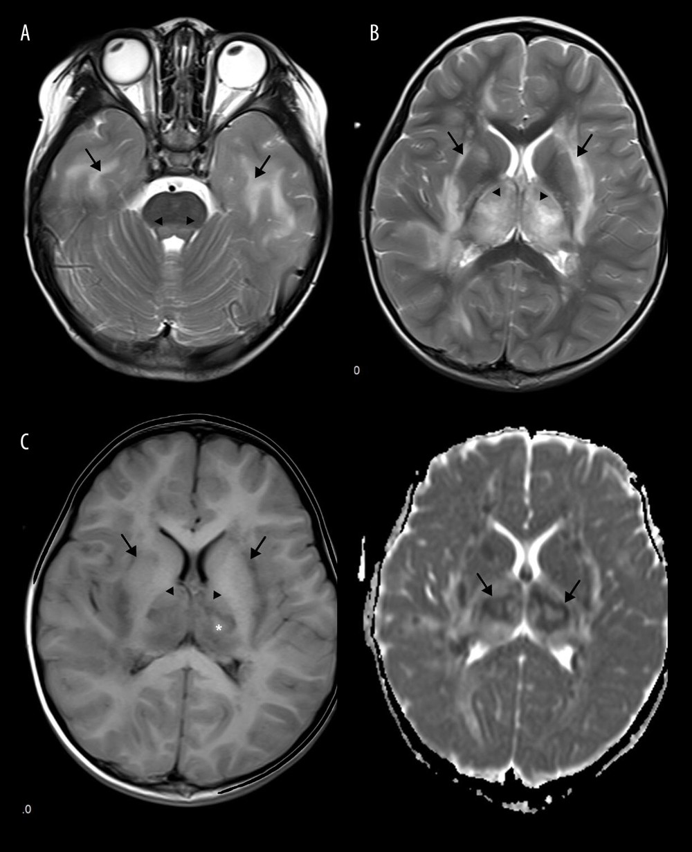
Figure 5 Emesia, febris for 5 days, followed by coma, with throat wrap for influenza type B positive (A–C). (A) Axial T2WI MRI showed mildly higher focal signal intensity in the dorsal aspect of pons (arrow), and mildly higher patchy signal intensity in white matter at bilateral temporal lobe (arrow). (B) Axal T2WI MRI indicated symmetric swelling and higher signal intensity in bilateral thalami (arrow), and white matter in external capsule was involved. (C) Axial T1WI MRI indicated mildly lower signal intensity in bilateral thalami (arrow) with slightly higher signal intensity in center (*), involvement of white matter in external capsule (*) was visible; axial ADC map showed more detailed 3-layer structure of bilateral thalami in ANE (arrow).