24 April 2021: Clinical Research
Anatomy and Correlation of the Coracoid Process and Coracoclavicular Ligament Based on Three-Dimensional Computed Tomography Reconstruction and Magnetic Resonance Imaging
Lan Xin 1ABCDEFG* , Jin Luo 2BF* , Mingying Chen 3AE* , Bing He 1AC* , Bi Tang 1BD* , Chunyang Tang 1DF , Dongyu Zhang 1BC , Lei Zhang 4567ABCF*DOI: 10.12659/MSM.930435
Med Sci Monit 2021; 27:e930435
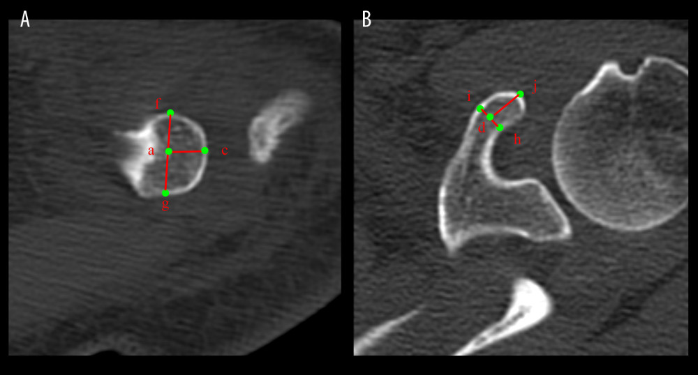
Figure 2 Image correlation data for computed tomography of the coracoclavicular (CC) ligament. (A) Subclavian observation. (B) superior coracoid process observation. Point a: The center of the CC ligament at the clavicular attachment. Point c: The farthest point at the acromion end of the clavicle. Point d: The point of the CC ligament at the center of the clavicular attachment. Point f: The point of the CC ligament at the anterior edge of the clavicular attachment. Point g: The posterior margin of the CC ligament at the clavicle attachment. Point h: The anterior edge of the CC ligament attached to the coracoid process. Point i: The posterior margin of the CC ligament attached to the coracoid process. Point j: Apical apex of the coracoid process. ac: Distance from the center point of the CC ligament at the supraclavicular attachment to the acromioclavicular joint. fg: The maximum diameter of the CC ligament at the anterior and posterior margin of the clavicle attachment. hi: The largest diameter of the CC ligament at the anterior and posterior edge of the coracoid process attachment. dj: The length of the CC ligament from the center point of the coracoid process attachment to the coracoid process tip.