24 April 2021: Clinical Research
Anatomy and Correlation of the Coracoid Process and Coracoclavicular Ligament Based on Three-Dimensional Computed Tomography Reconstruction and Magnetic Resonance Imaging
Lan Xin 1ABCDEFG* , Jin Luo 2BF* , Mingying Chen 3AE* , Bing He 1AC* , Bi Tang 1BD* , Chunyang Tang 1DF , Dongyu Zhang 1BC , Lei Zhang 4567ABCF*DOI: 10.12659/MSM.930435
Med Sci Monit 2021; 27:e930435
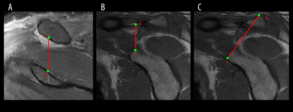
Figure 1 T2-weighted image correlation data for coracoclavicular (CC) ligament magnetic resonance imaging (MRI). (A) Measurement of the length of the CC ligament on T2-weighted MRI. (B) Sagittal coracoclavicular ligament length measurement on T2-weighted MRI. (C) Measurement of the distance from the supraclavicular plane to the subcoracoid plane on T2-weighted MRI. Point a: The central point of the CC ligament at the clavicular attachment. Point b: The CC ligament at the center of the CC attachment. Point d: The CC ligament at the center of the clavicular attachment. Point e: The CC ligament at the coracoid attachment point. Point k: The point of the supraclavicular plane through the CC ligament. Point l: The point of the subcoracoid plane through the CC ligament the length of the CC ligament. ab: The length of the CC ligament in the coronal plane. de: The length of the CC ligament in the sagittal plane. kl: The distance between the supraclavicular plane of the CC ligament and the subcoracoid process plane.