06 November 2020: Clinical Research
A Comparison of 2 Anterior Hybrid Techniques for 3-Level Cervical Degenerative Disc Disease
Han Wang BCE* , Yang Meng DF* , Hao Liu A* , Xiaofei Wang C , Chen Ding FDOI: 10.12659/MSM.927972
Med Sci Monit 2020; 26:e927972
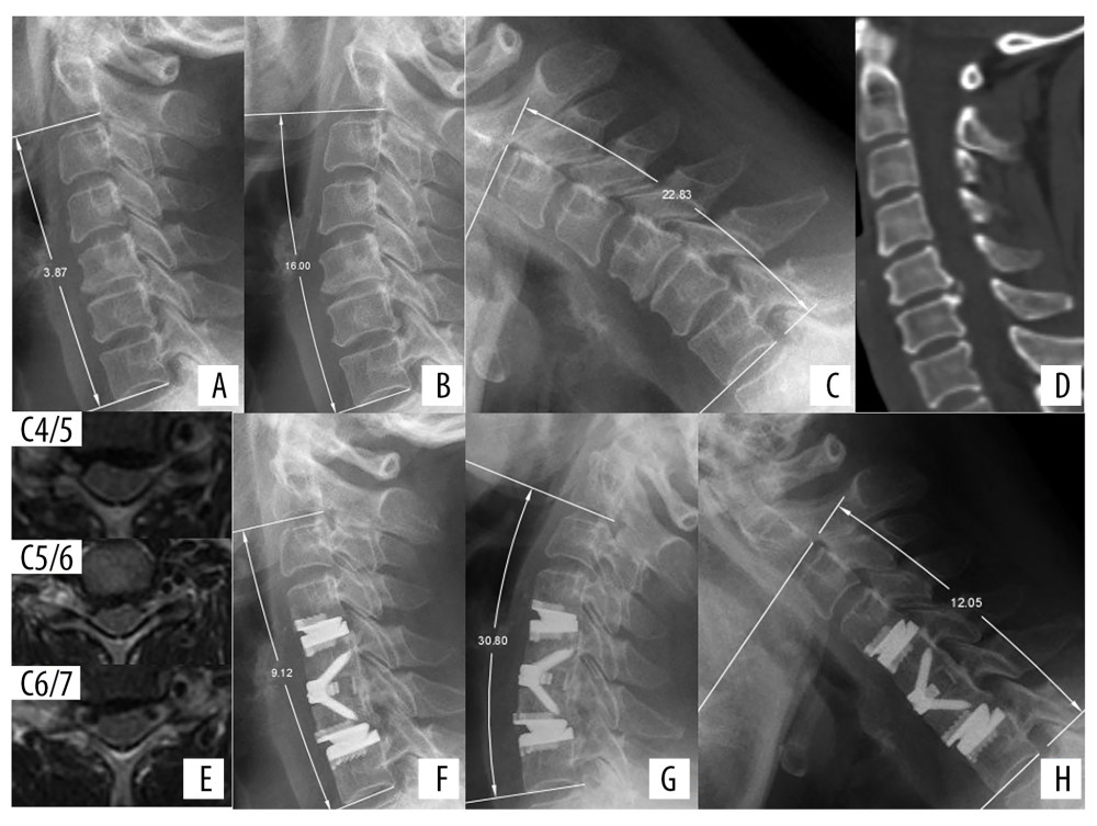
Figure 1 Radiologic examinations of a 55-year-old woman with neck pain for more than 2 years. (A) Preoperative lateral X-ray showing cervical lordosis at C2–C7 of 3.87°. (B, C) Flexion-extension view, showing that ROM at C2–C7 was 38.83°. (D) CT scan, showing osteophytes at the posterior border of C4–C5. (E) MRI showing herniated cervical discs at C4/5, C5/6, and C6/7, causing pressure on the spinal cord. CDR was performed at C4/5 and C6/7 and ACDF at C5/6. (F) X-ray immediately after surgery, showing a cervical lordosis of 9.12°. (G, H) Flexion-extension view at 1 year, showing that ROM at C2–C7 was 42.85° (110.35% compared with preoperative ROM).