21 October 2020: Clinical Research
Clinical Utility of 3-Dimensional Reconstruction Images to Predict Conservative Treatment Outcomes of Intra-Articular Distal Radius Fractures
Lingde Kong 1ABCF* , Zuzhuo Zhang 2BCD* , Jian Lu 1CDF , Bing Zhang 1EF , Yanqing Zhou 1BCD , Dehu Tian 1AEF*DOI: 10.12659/MSM.926894
Med Sci Monit 2020; 26:e926894
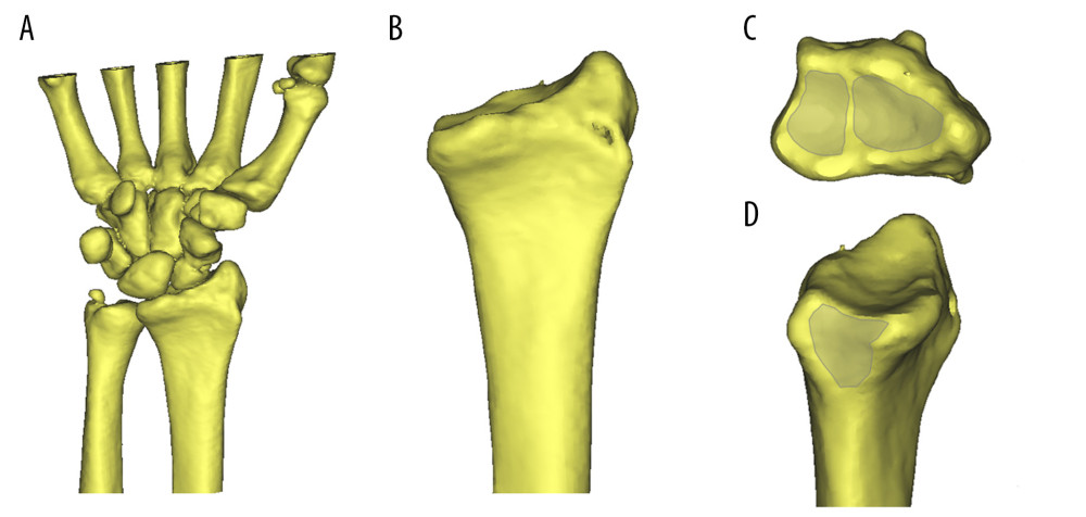
Figure 1 Three-dimensional (3D) images of articular surface were reconstructed by use of computed tomography images: (A) 3D images of wrist joint; (B) 3D images of distal radius; (C) scaphoid fossa and lunate fossa from the upper view; (D) sigmoid notch of distal radius from the ulnar view.