18 November 2020: Animal Study
Altered Inflammatory Pathway but Unaffected Liver Fibrosis in Mouse Models of Nonalcoholic Steatohepatitis Involving Interleukin-1 Receptor-Associated Kinase 1 Knockout
Ying Lei BCDE* , Tianxiao Yang BEFG* , Aijing Shan CDG , Wei Di BC , Mengyao Dai EFG , Jingminjie Nan B , Dongxue Liu B , Yanan Cao AEG , Xiuli Jiang AEG*DOI: 10.12659/MSM.926187
Med Sci Monit 2020; 26:e926187
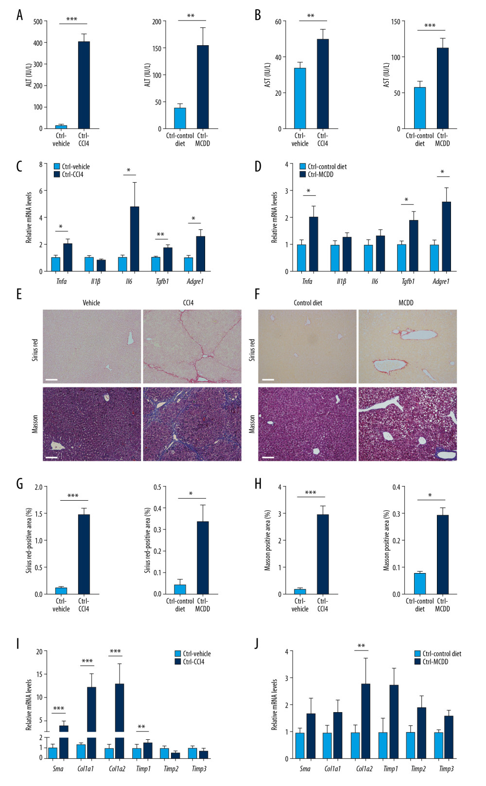
Figure 2 Generation of NASH in mice. (A, B) Serum (A) ALT and (B) AST levels of control mice treated for 8 weeks with CCl4 or vehicle (n=4–6) or fed MCDD or a control diet for 6 weeks (n=5). (C, D) Hepatic Tnfα, Il1β, Il6, Tgfb1, and Adgre1 mRNA levels in control mice (C) treated for 8 weeks with CCl4 or vehicle (n=4–6) or (D) fed MCDD or a control diet for 6 weeks (n=5). (E, F)4Representative images of Sirius red (upper part) and Masson staining (bottom part) of hepatic tissues of control mice (E) treated for 8 weeks with CCl4 or vehicle (n=4–6) or (F) fed MCDD or a control diet for 6 weeks (n=3–4). Scale bars, 100 μm. (G, H) Digital quantification of (G) Sirius red-positive and (H) Masson stain-positive areas in control mice treated for 8 weeks with CCl4 or vehicle (n=4–6) or fed MCDD or a control diet for 6 weeks (n=3–4). (I, J) Hepatic Sma, Col1a1, Col1a2, Timp1, Timp2, and Timp3 mRNA levels in control mice (I) treated for 8 weeks with CCl4 or vehicle (n=4–6) or (J) fed MCDD or a control diet for 6 weeks (n=5). Data represent mean±SEM. * p<0.05, ** p<0.01, *** p< 0.001, by t test.