23 November 2020: Clinical Research
Altered Brain Network Centrality in Patients with Adult Strabismus with Amblyopia: A Resting-State Functional Magnetic Resonance Imaging (fMRI) Study
Kang-Rui Wu 1ABCD* , Ya-Jie Yu 1ABCD* , Li-Ying Tang 2BCDE* , Si-Yi Chen 1BCDF , Meng-Yao Zhang 1ABCD , Tie Sun 1ABCF , Shi-Nan Wu 1ABCF , Kang Yu 1BCDF , Biao Li 1BCF , Yi Shao 1ABCDEF*DOI: 10.12659/MSM.925856
Med Sci Monit 2020; 26:e925856
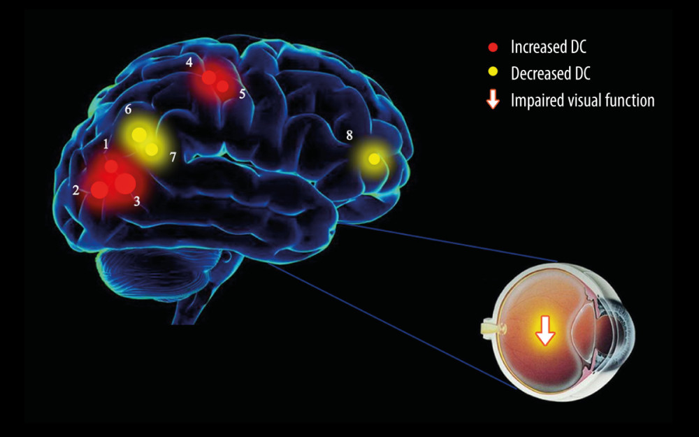
Figure 4 The DC results of brain activity in the SA group. Compared with the HCs, the DC of the following regions were increased to various extents: 1 – right lingual gyrus (t=4.6259); 2 – right middle occipital gyrus (t=5.6222); 3 – left fusiform gyrus (t=7.6374); 4 – left paracentral lobule (t=5.1415); 5 – right postcentral gyrus (t=4.1174); 6 – right angular gyrus (t=−5.2001); 7 – left angular gyrus (t=−4.439); 8 – left middle frontal gyrus (t=−4.0959) in SA patients. The sizes of the spots denote the degree of quantitative changes. DC – degree centrality; SA – strabismus with amblyopia; HC – healthy control.