Logo Medical Science Monitor

Call: +1.631.470.9640
Mon - Fri 10:00 am - 02:00 pm EST

Contact Us

Logo Medical Science Monitor Logo Medical Science Monitor Logo Medical Science Monitor

12 October 2020: Clinical Research

Three-Dimensional Computed Tomography (CT) Mapping of Intertrochanteric Fractures in Elderly Patients

Cong Li 1ACEF , Dongyang Zhao 1BE , Xian Xu 1B , Jiajun Ding 1F , Yanping Guo 1F , Lili Liao 2AD** , Guang Li 1BCD**

DOI: 10.12659/MSM.925452

Med Sci Monit 2020; 26:e925452

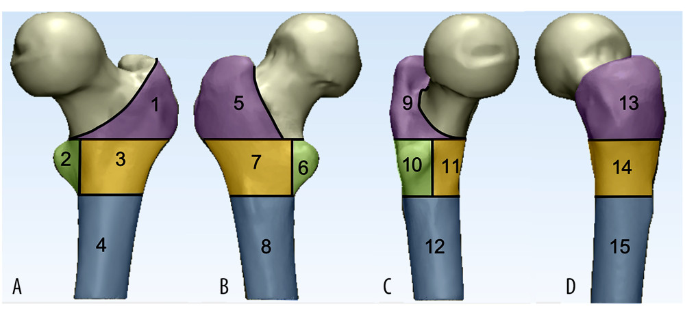
Figure 2 Osseous zone models. (A) Anteroposterior, (B) posteroanterior, (C) medial, medial, (D) medial. Several separate lines divide the proximal femur into 4 osseous zones: greater trochanter area (purple area), lesser trochanter area (reseda area), lesser trochanter lateral area (yellow area), subtrochanteric area (wathet area).

Your Privacy

We use cookies to ensure the functionality of our website, to personalize content and advertising, to provide social media features, and to analyze our traffic. If you allow us to do so, we also inform our social media, advertising and analysis partners about your use of our website, You can decise for yourself which categories you you want to deny or allow. Please note that based on your settings not all functionalities of the site are available. View our privacy policy.

Medical Science Monitor eISSN: 1643-3750